. 2021 Jun;74(6):1429-1441.
doi: 10.1016/j.jhep.2021.01.028. Epub 2021 Jan 27.

STARD1 promotes NASH-driven HCC by sustaining the generation of bile acids through the alternative mitochondrial pathway

Affiliations

STARD1 promotes NASH-driven HCC by sustaining the generation of bile acids through the alternative mitochondrial pathway

Laura Conde de la Rosa et al. J Hepatol. 2021 Jun.

Abstract

Background & aims: Besides their physiological role in bile formation and fat digestion, bile acids (BAs) synthesised from cholesterol in hepatocytes act as signalling molecules that modulate hepatocellular carcinoma (HCC). Trafficking of cholesterol to mitochondria through steroidogenic acute regulatory protein 1 (STARD1) is the rate-limiting step in the alternative pathway of BA generation, the physiological relevance of which is not well understood. Moreover, the specific contribution of the STARD1-dependent BA synthesis pathway to HCC has not been previously explored.

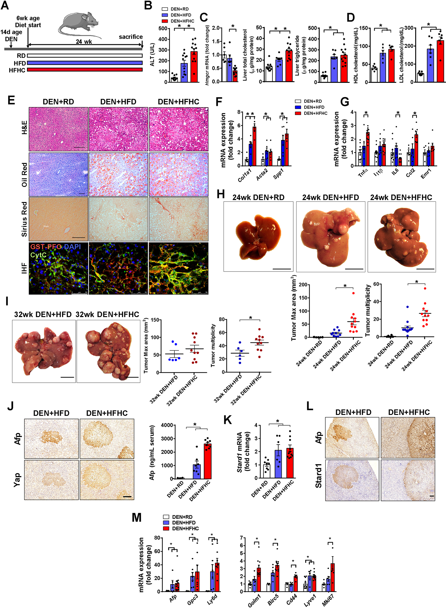
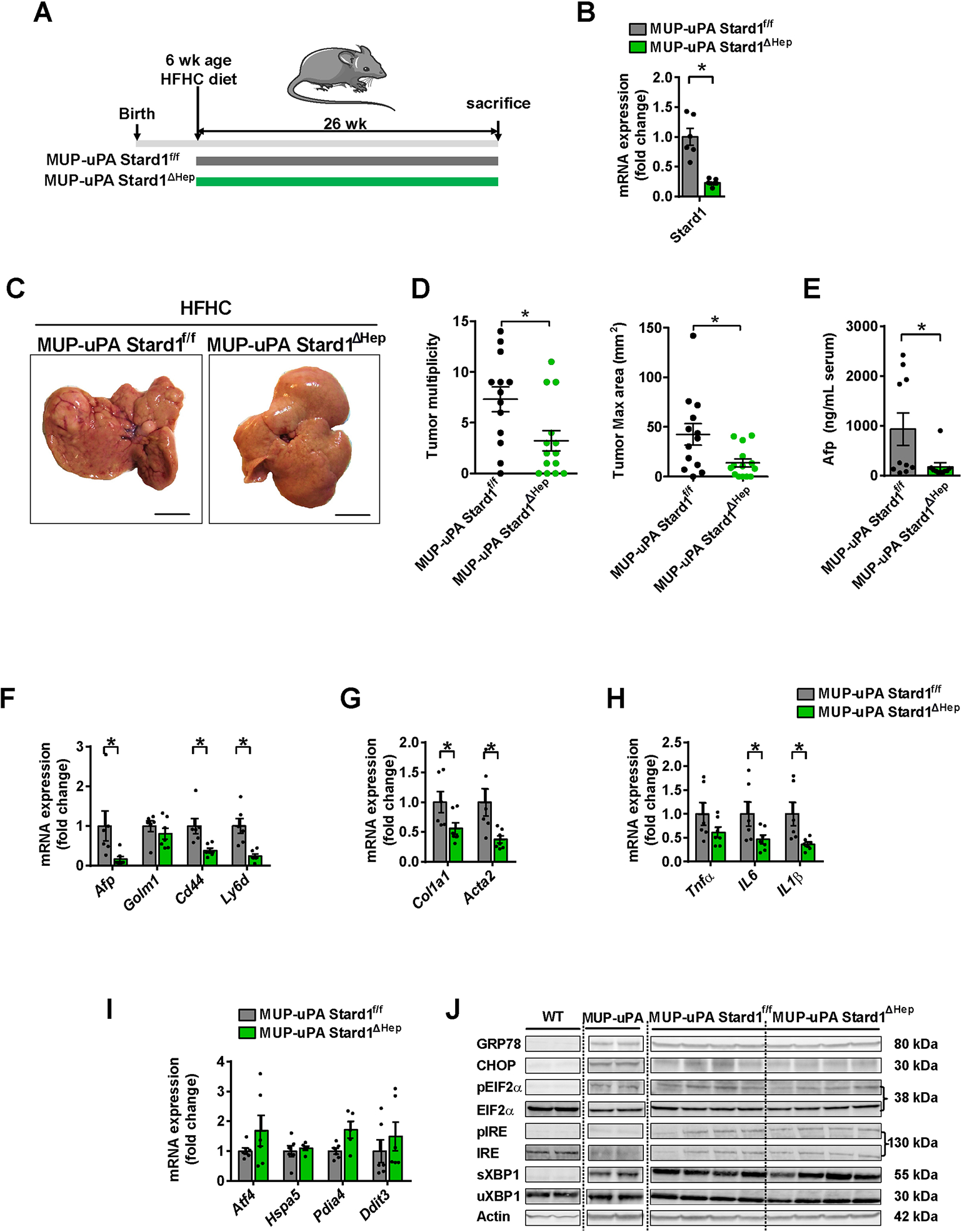
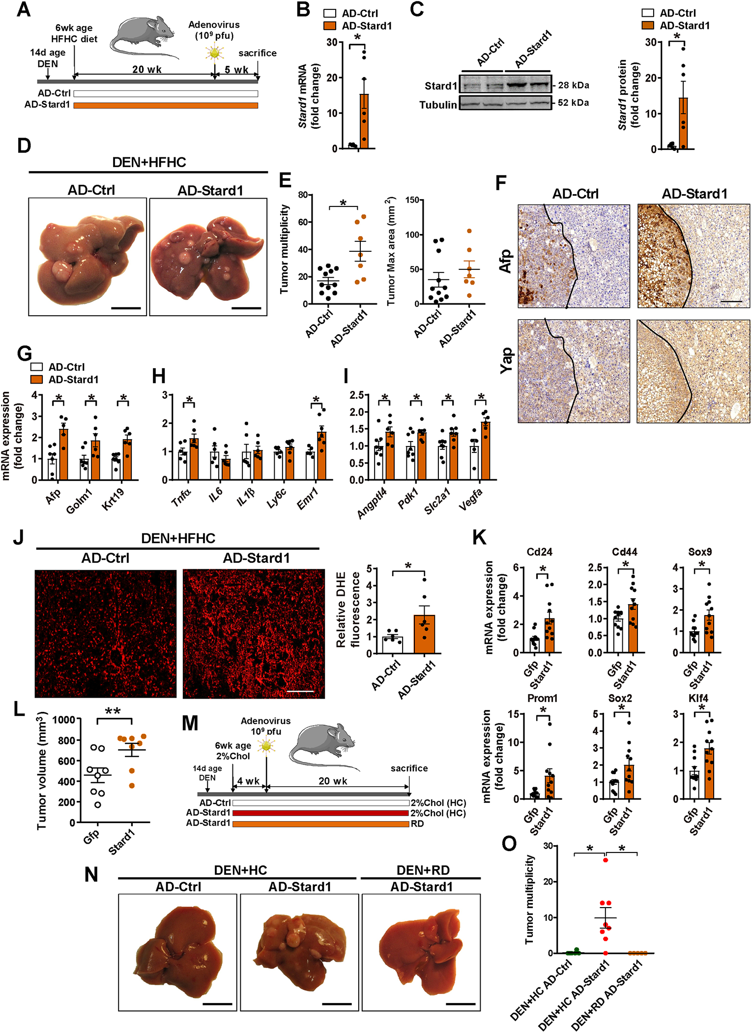
Methods: STARD1 expression was analyzed in a cohort of human non-alcoholic steatohepatitis (NASH)-derived HCC specimens. Experimental NASH-driven HCC models included MUP-uPA mice fed a high-fat high-cholesterol (HFHC) diet and diethylnitrosamine (DEN) treatment in wild-type (WT) mice fed a HFHC diet. Molecular species of BAs and oxysterols were analyzed by mass spectrometry. Effects of NASH-derived BA profiles were investigated in tumour-initiated stem-like cells (TICs) and primary mouse hepatocytes (PMHs).

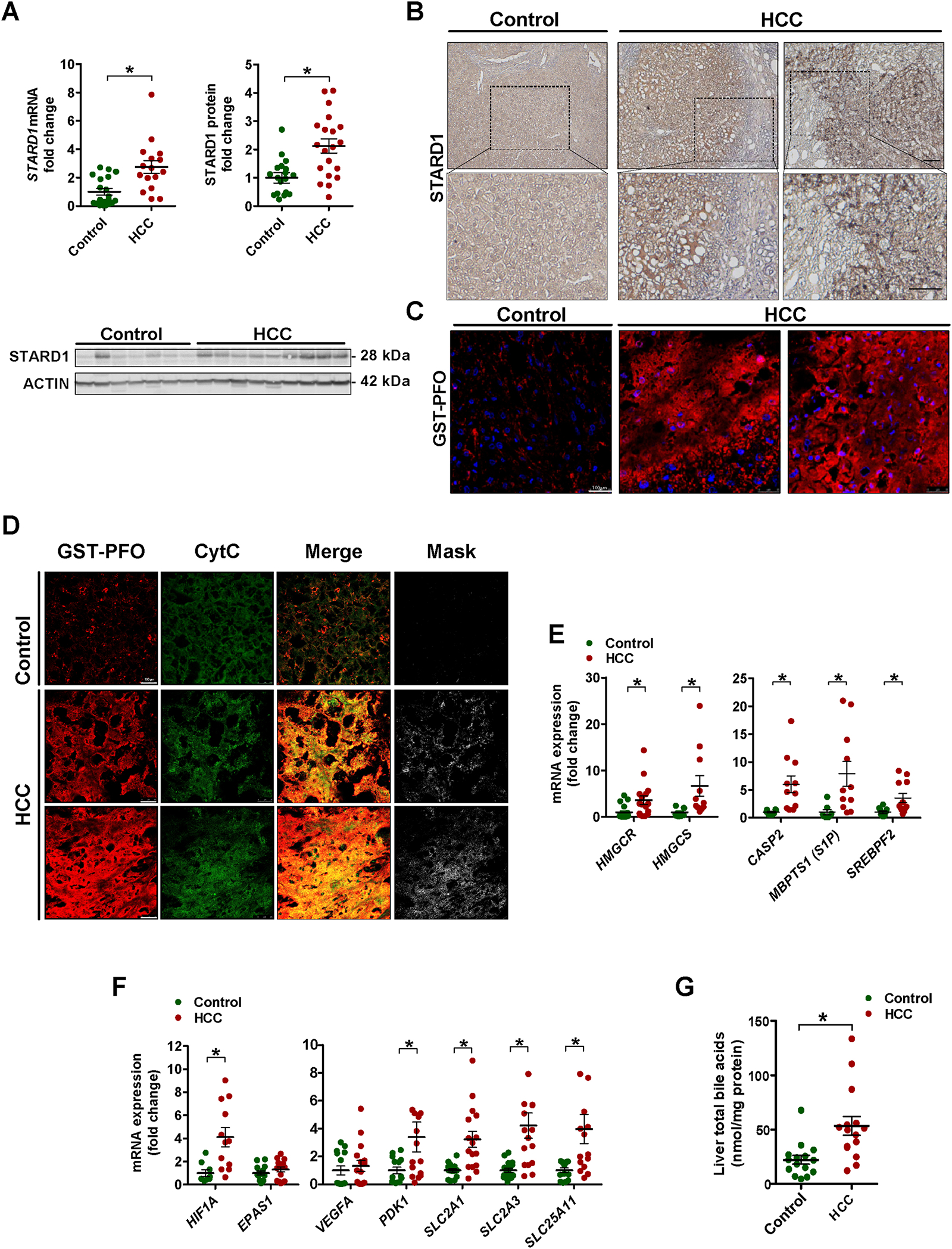
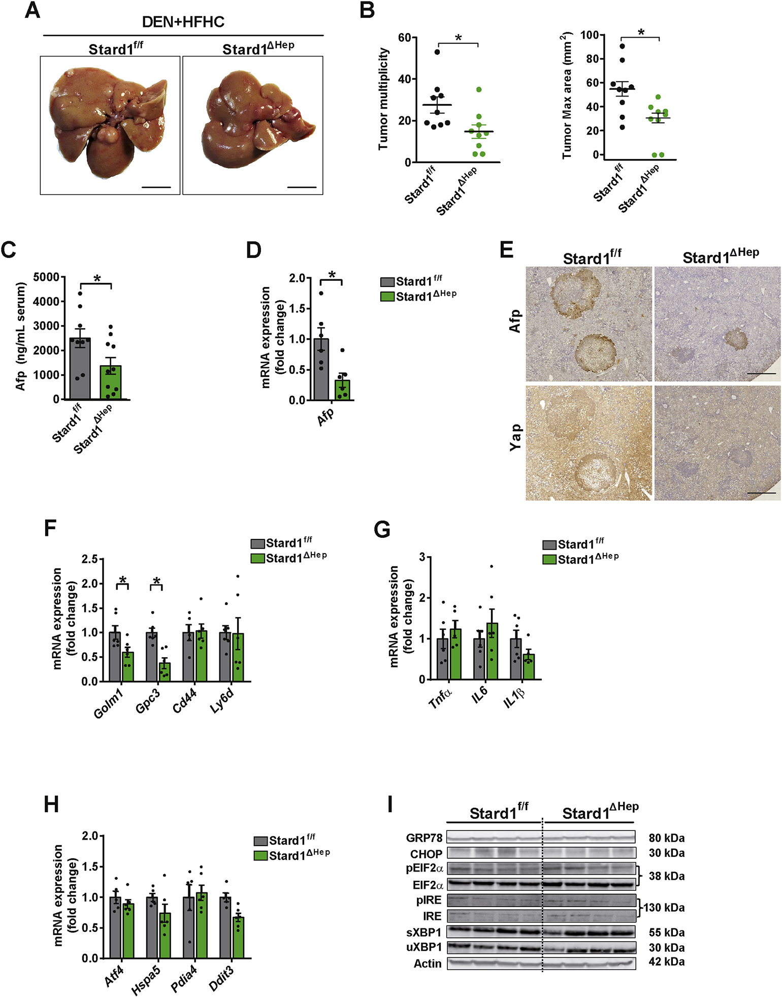
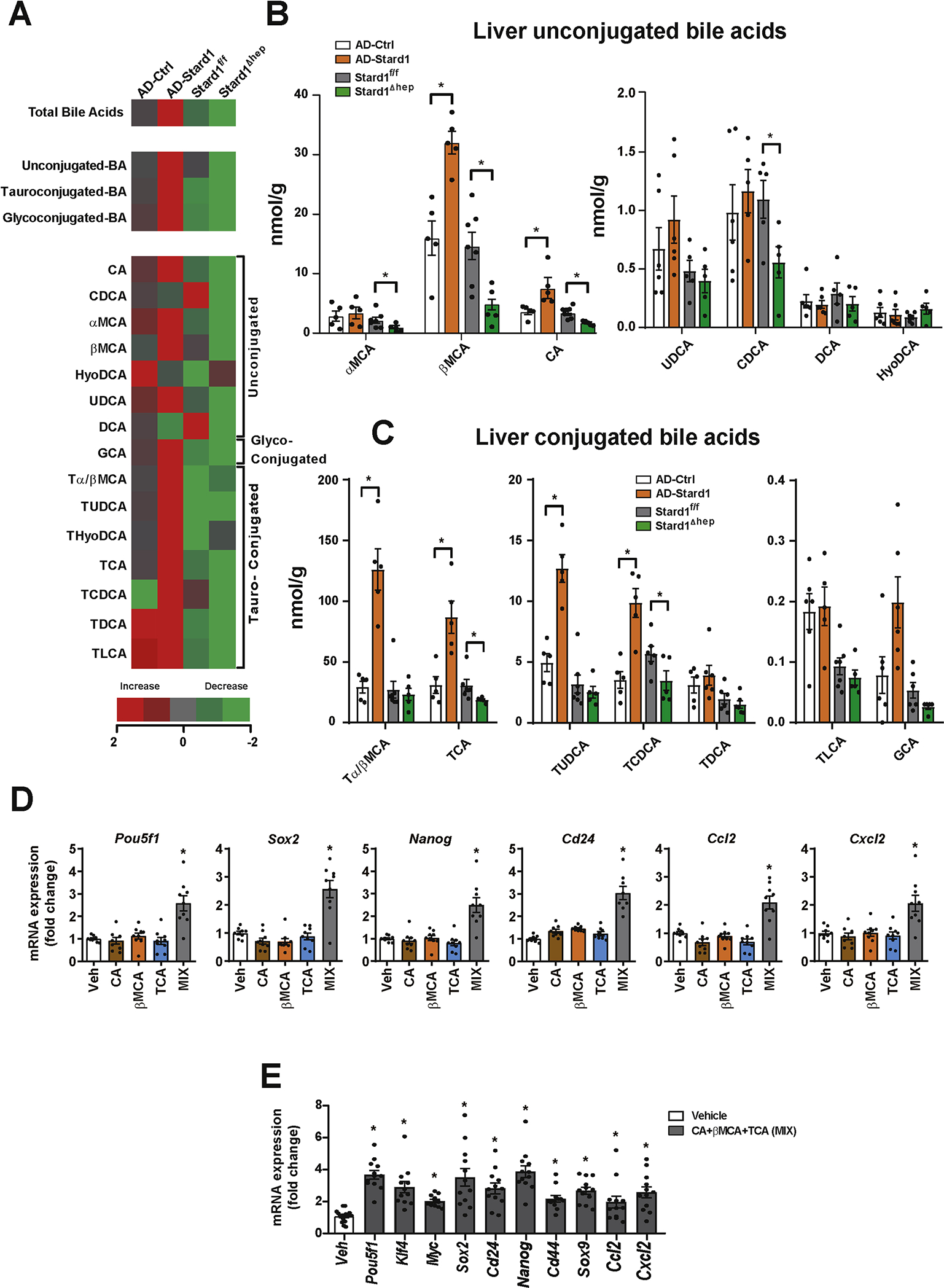
Results: Patients with NASH-associated HCC exhibited increased hepatic expression of STARD1 and an enhanced BA pool. Using NASH-driven HCC models, STARD1 overexpression in WT mice increased liver tumour multiplicity, whereas hepatocyte-specific STARD1 deletion (Stard1ΔHep) in WT or MUP-uPA mice reduced tumour burden. These findings mirrored the levels of unconjugated primary BAs, β-muricholic acid and cholic acid, and their tauroconjugates in STARD1-overexpressing and Stard1ΔHep mice. Incubation of TICs or PMHs with a mix of BAs mimicking this profile stimulated expression of genes involved in pluripotency, stemness and inflammation.

Conclusions: The study reveals a previously unrecognised role of STARD1 in HCC pathogenesis, wherein it promotes the synthesis of primary BAs through the mitochondrial pathway, the products of which act in TICs to stimulate self-renewal, stemness and inflammation.

Lay summary: Effective therapy for hepatocellular carcinoma (HCC) is limited because of our incomplete understanding of its pathogenesis. The contribution of the alternative pathway of bile acid (BA) synthesis to HCC development is unknown. We uncover a key role for steroidogenic acute regulatory protein 1 (STARD1) in non-alcoholic steatohepatitis-driven HCC, wherein it stimulates the generation of BAs in the mitochondrial acidic pathway, the products of which stimulate hepatocyte pluripotency and self-renewal, as well as inflammation.

Keywords: Bile acids; Cholesterol; Hepatocellular carcinoma; Mitochondria; Oxysterols; STARD1.


Conflict of interest statement

Conflicts of interest The authors declare no conflicts of interest that pertain to this work. Please refer to the accompanying ICMJE disclosure forms for further details.

Figures

Figure 1.
Figure 1.. Increased STARD1 expressed in human NASH-driven HCC.
A) Expression of STAR gene mRNA by qPCR and STARD1 protein in liver tissue from control donors and NASH-driven HCC (N of controls =15, N of HCC patients 15–20). B) Representative immunohistochemical expression of STARD1 from control and NASH-HCC patient liver samples. C) Staining of liver sections from control and HCC samples with GST-PFO (red) to detect free cholesterol. Nuclei were stained with DAPI. D) Immunostaining of liver sections from control and HCC samples with GST-PFO and cytochrome c (Cyt C), showing their colocalization as merge and mask. Bar 75 μm. E) Transcript quantification by qPCR of genes controlling cholesterol biosynthesis, ER stress-driven activation of SREBP2. F) mRNA levels of HIF1A and HIF2A (EPAS1) and HIF-1α regulated genes. N of control 13–18, N of HC 13–20. G) Hepatic levels of total BAs in samples from control and human HCC. (N=15 both groups). All values are mean ± SEM. * indicate statistically significant differences between the indicated groups (p<0.05) in Student’s t test. Magnification bar in histology pictures, 100 μm.
Figure 2.
Figure 2.. HFHC feeding promotes NASH-driven HCC development in DEN-treated wild type.
A) Schematic illustration of the experimental design, with induction of tumorogenesis in liver of mice with DEN at 14 days of age, feeding with regular diet (RD), high fat diet (HFD) or cholesterol-supplemented HFD (HFHC) diet for 24 weeks. N per group: RD (11), HFD (12), HFHC (12). B) Transaminase serum levels (ALT) of mice after the corresponding treatments. C) Liver Hmgcr transcript, total cholesterol and triglycerides liver composition. N=6 per group. D) HDL and LDL levels in serum from DEN+HFD or DEN+HFHC fed mice. N=6 per group. E) Representative histological staining for hematoxylin-eosin (H&E), neutral lipid (oil red o) and collagen fibers (sirius red) of liver sections. Immunohistofluorescence of liver sections stained for free cholesterol with GST-PFO probe (red), mitochondria with anti-cytochrome c (green) and nuclei with Dapi (blue). Size bar 100 μm. F) mRNA levels of fibrogenesis-associated genes (Col1a1, Acta2, Spp1). All values are corrected by a housekeeping gene (Actb) and relative to values from the animals of DEN- RD diet. N=6–10 per group. G) mRNA levels of inflammation genes (Tnfa, Il1b, Il6, Ccl2, Emr1). H) Representative macroscopic images and quantification of tumor multiplicity and maximal area from DEN-treated mice fed HFHC diet for 24 weeks. RD, N=6; HFD, N=10; HFHC, N=11. I) As in H) except that DEN-treated mice were fed HFHC diet for 32 weeks. HFD, N=6; HFHC, N=10. J) Immunohistochemical expression of Afp and Yap of liver consecutive sections from DEN-treated mice fed HFC or HFHC diet for 24 weeks. K) mRNA levels of Stard1 of whole liver tissue from DEN-treated mice fed RD or HFHC diet. N=6–10 per group. L) Immunohistochemistry of consecutive sections (T, tumor) stained for Afp, or Star. Size bar, 500 μmeter. M) mRNA levels of tumor markers and inflammatory genes of whole liver tissue from DEN-treated mice fed RD, HFD or HFHC. N=6–10 per group. All values are mean ± SEM; symbol * indicates statistically significant differences (p<0.05) on a one-way ANOVA test or student’s t test.
Figure 3.
Figure 3.. Hepatocyte Stard1 deletion in MUP-uPA mice attenuates NASH-driven HCC in mice.
A) Feeding of MUP-uPA Stard1f/f and MUP-uPA Stard1 ΔHep mice with HFHC diet for 26 weeks. B) mRNA levels of Stard1 in MUP-uPA Stard1f/f (N=6) and MUP-uPA Stard1 ΔHep mice (N=7). C-D) Macroscopic images of livers from MUP-uPA Stard1f/f (N=13) and MUP-uPA Stard1 ΔHep mice (N=14) fed HFHC diet for 26 weeks, with quantification of tumor multiplicity and maximal area. E) Serum Afp levels of MUP-uPA Stard1f/f (N=10) and MUP-uPA Stard1 ΔHep mice (N=10) fed HFHC diet. F-H) mRNA levels tumor markers, fibrosis and inflammation genes of whole liver tissue from MUP-uPA Stard1f/f (N=6) and MUP-uPA Stard1 ΔHep mice (N=7) fed HFHC. I) mRNA levels of ER stress markers of whole liver tissue from MUP-uPA Stard1f/f (N=6) and MUP-uPA Stard1 ΔHep mice (N=7) fed HFHC. J) Western blot of ER stress markers as in H). All values are mean ± SEM. *p<0.05, denote statistically significant differences respect to MUP-uPAStard1f/f or Stard1f/f mice in Student’s t test.
Figure 4.
Figure 4.. Stard1 ΔHep mice are less sensitive to DEN plus HFHC induced HCC.
A-B) Macroscopic images of livers from Stard1f/f (N=9) and Stard1ΔHep mice (N=9) treated with DEN and fed HFHC diet for 24 weeks, with quantification of tumor multiplicity and maximal area. C-D) Serum and mRNA expression levels of Afp from Stard1f/f (N=9) and Stard1ΔHep mice (N=9) treated with DEN and fed HFHC. E) Immunohistochemical expression of Afp and Yap of consecutive liver sections from Stard1f/f and Stard1 ΔHep mice. F-G) mRNA levels tumor markers and inflammation genes of whole liver tissue from Stard1f/f and Stard1ΔHep mice. H) mRNA levels of ER stress markers of whole liver tissue from Stard1f/f (N=6) and Stard1ΔHep mice (N=6). I) Western blot of ER stress markers as in H). All values are mean ± SEM. *p<0.05, denote statistically significant differences respect to MUP-uPAStard1f/f or Stard1f/f mice in Student’s t test.
Figure 5.
Figure 5.. Stard1 overexpression increases DEN+HFHC-driven HCC.
A) Schematic illustration of the experimental design used to overexpress Stard1 in wild type mice 5 months after DEN+HFHC treatment. B) Adenoviral-induced overexpression of mouse Stard1 in liver of mice determined by qPCR. N=6 for each group. C) Quantification of Stard1 overexpression by immunoblot densitometry and representative image of a Western blot for Stard1. N=6 for each group. D-E) Representative images of livers and quantification of macroscopic liver tumor multiplicity and maximum size in animals after 5 weeks of recombinant adenovirus injection. N=11 for AD-Ctrl and N=7 for AD-Stard1. F) Immunohistochemistry of consecutive sections showing the same tumor (T, delimited with a dotted line) and parenchyma stained for Afp, or Yap. Size bar 500 μmeter. G, H, I) qPCR quantification of mRNA of HCC markers, fibrogenesis, inflammation and Hif1a target genes. N=6 per group. J) ROS production measured and quantified in cryosections of liver tissue stained with DHE, (N=6). K) mRNA levels of stemness genes measured in subcutaneous tumors induced by TICs with or without Stard1 overexpression. Values are mean ± SEM relative to the Gfp-expressing tumors (N=12). * denotes statistically significance in paired Student’s t test respect the matched Gfp-expressing tumors. L) Subcutaneous tumor volume in nude mice induced by TICs transfected with control Gfp or Stard1. Values are mean ± SEM relative to the Gfp-expressing tumors (N=8). * denotes statistically significance in paired Student’s t test respect the matched Gfp-expressing tumors. M) Schematic experimental design for adenoviral-mediated Stard1 overexpression (AD-Star) in DEN-treated wild type mice followed by feeding a regular diet (RD) (N=5) or a diet enriched in cholesterol (2%, HC) (N=8) or AD-Ctrl on HC diet (N=6). N, O) Macroscopic images of livers from DEN-treated mice and quantification of tumor multiplicity. All values are mean ± SEM. *p<0.05 denote statistically significant differences respect to Ad-Ctrl in a student’s t test.
Figure 6.
Figure 6.. Molecular species of BAs in NASH-HCC models and their impact in expression of genes involved in self-renewal, stemness and inflammation.
A) Heatmap of individual species of BAs measured in livers from AD-Stard1 mice and Stard1 ΔHep mice following DEN treatment and HFHC feeding, showing an increase (red) or a reduction (green) respect to the mean of AD-control and Stard1f/f mice. Values are Log2 of the fold change. B) Quantification of the unconjugated BAs in liver tissue from each group of animals. (N=5 for AD-Ctrl/AD-Stard1 and N=6 for Stard1f/f and Stard ΔHep mice. C) Quantification of tauroconjugated BAs in liver tissue from each group of animals. D) mRNA levels of genes involved in self-renewal, stemness and inflammation in TICs following incubation with CA (50 μM), βMCA (50 μM) and TCA (200 μM) for 48 hours. Values are mean ± SEM. N=3 independent experiments performed in triplicates. E) Effect of the combination of CA, βMCA and TCA in PMH for 24 hours on the mRNA levels of genes involved in self-renewal, stemness and inflammation. Values are mean ± SEM. N=3 independent experiments performed in quadruplicates.

Comment in

References

    1. Bianchini F, Kaaks R, Vainio H. Overweight, obesity, and cancer risk. The lancet oncology 2002;3:565–574. - PubMed
    1. Calle EE, Rodriguez C, Walker-Thurmond K, Thun MJ. Overweight, obesity, and mortality from cancer in a prospectively studied cohort of U.S. adults. The New England journal of medicine 2003;348:1625–1638. - PubMed
    1. Forner A, Reig M, Bruix J. Hepatocellular carcinoma. Lancet 2018;391:1301–1314. - PubMed
    1. Bruix J, Reig M, Sherman M. Evidence-Based Diagnosis, Staging, and Treatment of Patients With Hepatocellular Carcinoma. Gastroenterology 2016;150:835–853. - PubMed
    1. Villanueva A, Hernandez-Gea V, Llovet JM. Medical therapies for hepatocellular carcinoma: a critical view of the evidence. Nature reviews Gastroenterology & hepatology 2013;10:34–42. - PubMed

Publication types

MeSH terms

Substances