

Histologic validation of optical coherence tomography-based three-dimensional morphometric measurements of the human optic nerve head: Methodology and preliminary results
- PMID: 33516762
- PMCID: PMC8044038
- DOI: 10.1016/j.exer.2021.108475
Histologic validation of optical coherence tomography-based three-dimensional morphometric measurements of the human optic nerve head: Methodology and preliminary results
Abstract
Purpose: To compare the three-dimensional (3D) morphology of the deep load-bearing structures of the human optic nerve head (ONH) as revealed in vivo by spectral domain optical coherence tomography (SDOCT) with ex vivo quantitative 3D histology.
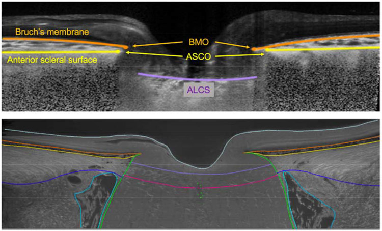
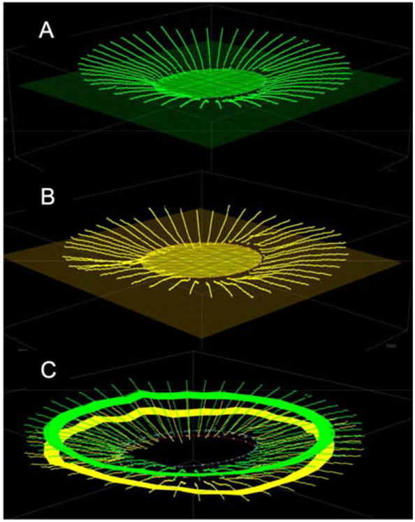
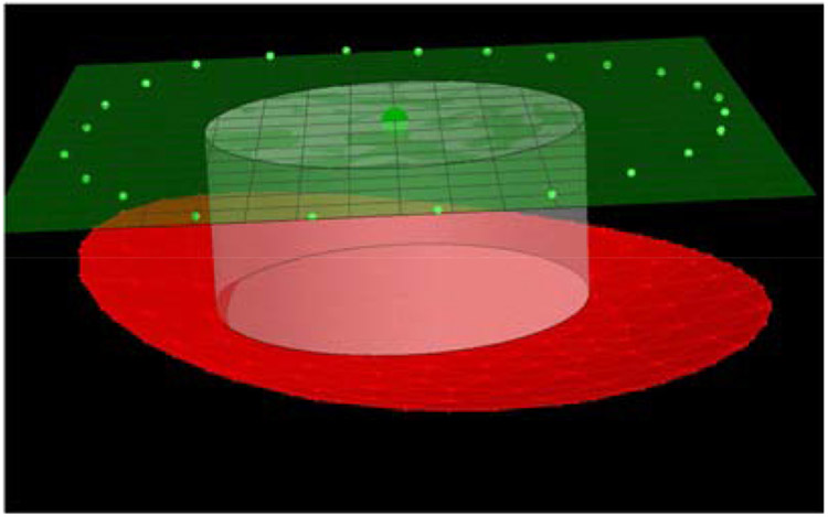
Methods: SDOCT imaging of the ONH was performed in six eyes from three brain-dead organ donors on life-support equipment awaiting organ procurement (in vivo conditions). Following organ procurement (ex vivo conditions), the eyes were enucleated and underwent a pars plana vitrectomy followed by pressurization to physiologic IOP and immersion fixation. Ex vivo ONH morphology was obtained from high-fidelity episcopic fluorescent 3D reconstruction. Morphologic parameters of the observed ONH canal geometry and peripapillary choroid, as well as the shape, visibility and depth of the lamina cribrosa were compared between ex vivo and in vivo measurements using custom software to align, scale, and manually delineate the different regions of the ONH.
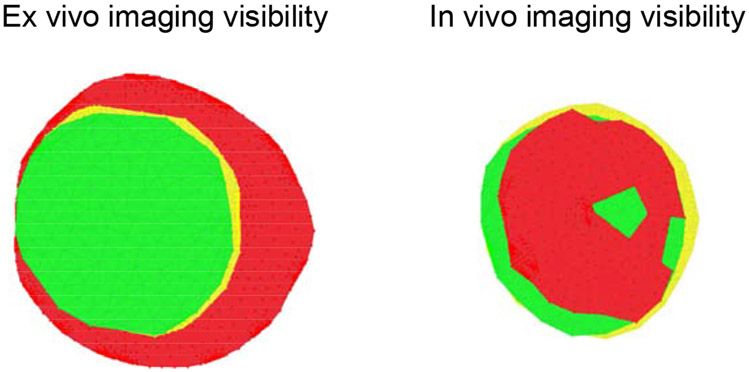
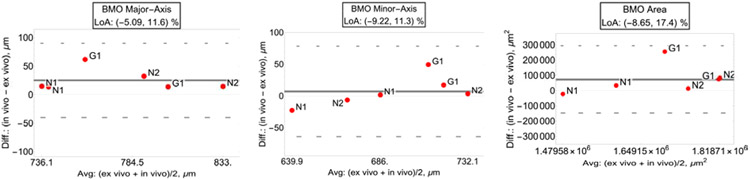
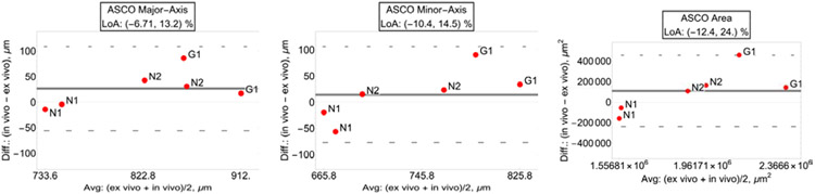
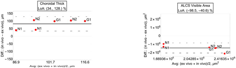
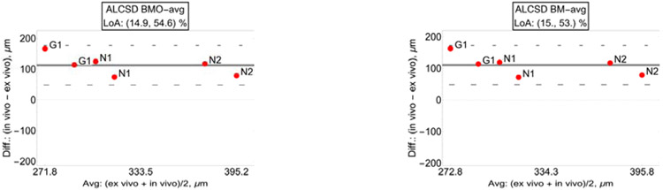
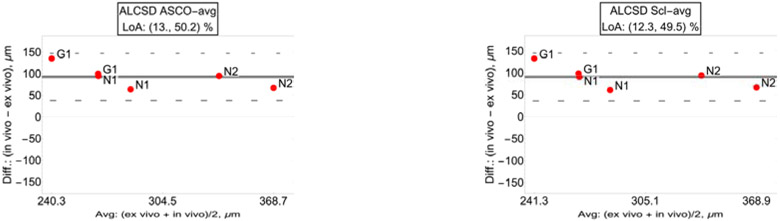
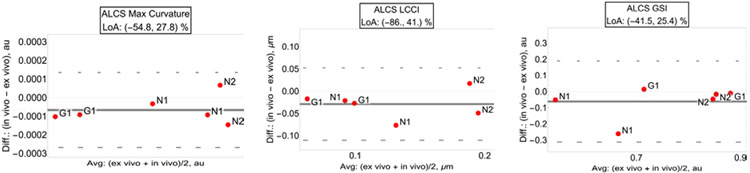
Results: There was significant correspondence between in vivo and ex vivo measurements of the depth and shape of the lamina cribrosa, along with the size and shape of Bruch's membrane opening (BMO) and anterior scleral canal opening (ASCO). Weaker correspondence was observed for choroidal thickness; as expected, a thinner choroid was seen ex vivo due to loss of blood volume upon enucleation (-79.9%, p < 0.001). In addition, the lamina was shallower (-32.3%, p = 0.0019) and BMO was smaller ex vivo (-3.38%, p = 0.026), suggesting post mortem shrinkage of the fixed tissue. On average, while highly variable, only 31% of the anterior laminar surface was visible in vivo with SDOCT (p < 0.001).
Conclusions: Morphologic parameters by SDOCT imaging of the deep ONH showed promising correspondence to histology metrics. Small but significant shrinkage artifact, along with large effects of exsanguination of the choroid, was seen in the ex vivo reconstructions of fixed tissues that may impact the quantification of ex vivo histoarchitecture, and this should be considered when developing models and biomarkers based on ex vivo imaging of fixed tissue. Lack of visibly of most of the lamina surface in SDOCT images is an important limitation to metrics and biomarkers based on in vivo images of the ONH deep tissues.
Keywords: Glaucoma; Histology; Optic nerve head morphology and biomechanics; Spectral domain optical coherence tomography.
Copyright © 2021 Elsevier Ltd. All rights reserved.
Conflict of interest statement
Figures
References
-
- Belghith A, Girkin CA, Weinreb RN, Medeiros F, Bowd C, Liebmann JM, Fazio MA, Zangwill LM, 2017. The African Descent and Glaucoma Evaluation Study (ADAGES): Racial differences in the posterior displacement of the anterior laminar cribrosa surface depth. Investigative Ophthalmology & Visual Science 58, 4015–4015. - PubMed
-
- Bellezza AJ, Hart RT, Burgoyne CF, 2000. The optic nerve head as a biomechanical structure: initial finite element modeling. Invest Ophthalmol Vis Sci 41, 2991–3000. - PubMed
-
- Bellezza AJ, Rintalan CJ, Thompson HW, Downs JC, Hart RT, Burgoyne CF, 2003. Deformation of the lamina cribrosa and anterior scleral canal wall in early experimental glaucoma. Invest Ophthalmol Vis Sci 44, 623–637. - PubMed
-
- Bland JM, Altman DG, 2007. Agreement Between Methods of Measurement with Multiple Observations Per Individual. Journal of Biopharmaceutical Statistics 17, 571–582. - PubMed
Publication types
MeSH terms
Grants and funding
LinkOut - more resources
Full Text Sources
Other Literature Sources
Miscellaneous
