

doi: 10.1016/j.cgh.2021.01.044.
Epub 2021 Jan 29.
Hepatic Vasculopathy and Regenerative Responses of the Liver in Fatal Cases of COVID-19
Affiliations
- PMID: 33516952
- PMCID: PMC7844358
- DOI: 10.1016/j.cgh.2021.01.044
Item in Clipboard
Hepatic Vasculopathy and Regenerative Responses of the Liver in Fatal Cases of COVID-19
Clin Gastroenterol Hepatol.
2021 Aug.
Abstract
Severe acute respiratory syndrome coronavirus-2 (SARS-CoV-2) infects the nasopharynx and lungs and causes coronavirus disease-2019 (COVID-19). It may impact the heart, brain, kidney, and liver.1 Although functional impairment of the liver has been correlated with worse clinical outcomes, little is known about the pathophysiology of hepatic injury and repair in COVID-19.2,3 Histologic evaluation has been limited to small numbers of COVID-19 cases with no control subjects2,4 and demonstrated largely heterogeneous patterns of pathology.2,3.
Copyright © 2021 The Authors. Published by Elsevier Inc. All rights reserved.
Figures
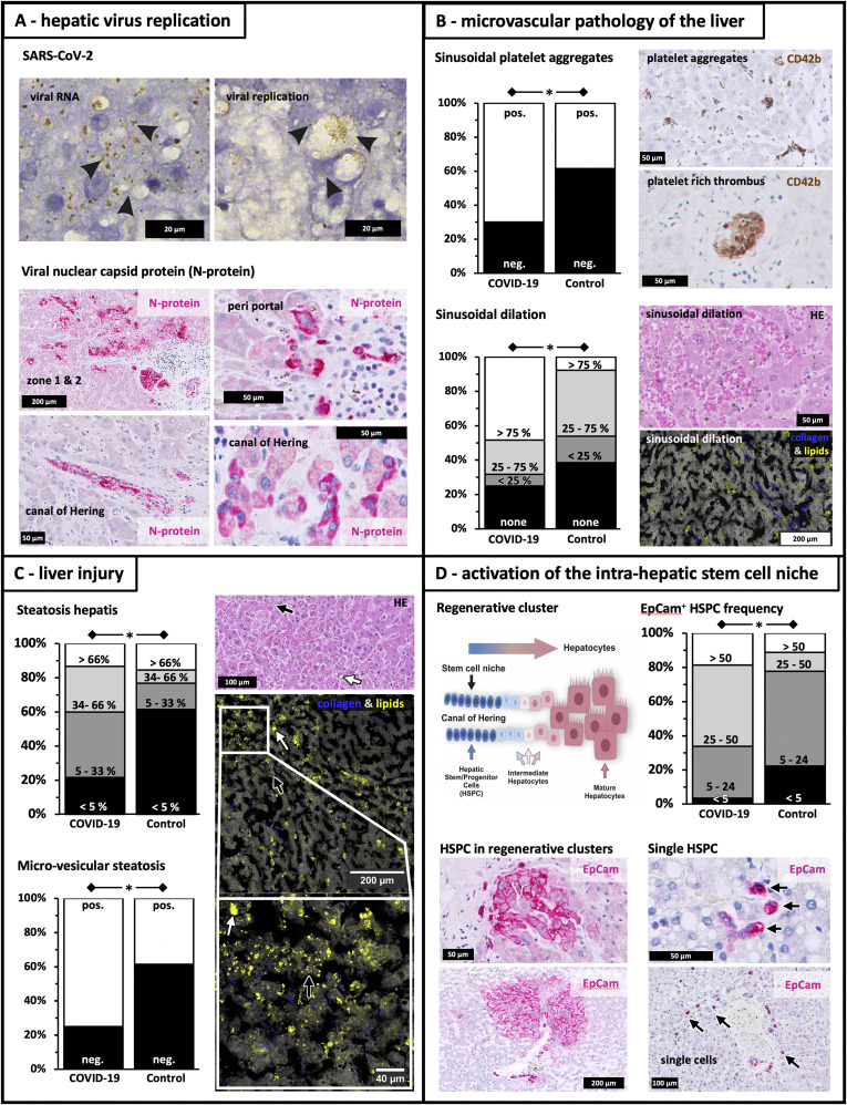
Microvascular pathology and regenerative responses in livers of COVID-19 patients. SARS-CoV-2 replication, histopathology, and regeneration responses of the liver in fatal COVID-19. (A) Viral replication. SARS-CoV-2 viral RNA (“viral RNA”; +strand, brown dots indicated by black arrows) detected by in situ hybridization and replicative viral intermediates (“viral replication”; -RNA, brown dots indicated by black arrows). Immunohistochemistry shows immunoreactivity against viral nucleocapsid protein (red) in hepatocytes within acinar zones I and II, periportal HSPC, cholangiocytes of portal bile ducts, and premature hepatocytes along the canal of Hering. (B) Microvascular pathology. Sinusoidal platelet aggregates and thrombi (anti-CD42b staining), frequent in COVID-19 patients. Sinusoidal dilatation is noted in hematoxylin and eosin (HE) staining. Coherent Raman scattering microscopy provided label-free contrast for tissue structure (grey) and lipid droplet identification (yellow), whereas second harmonic generation provided contrast for collagen fibers (blue). (C) Liver injury. High levels of hepatocyte steatosis in COVID-19 patients, in comparison with control subjects, as detected by HE staining and coherent Raman scattering/second harmonic generation (lipid droplets in yellow and collagen in blue). White arrows indicate macrovesicular steatosis, whereas black arrows show microvesicular steatosis. (D) Intrahepatic stem cell niche. Drawing of the bipotent intrahepatic stem cell compartment, which is located in the canal of Hering. On severe hepatic injury HSPC emerge and expand, contributing to liver repair. HSPC are detected as single cells (indicated by black arrows) or in regenerative clusters comprised of EpCAM+ HSPC, hepatobiliary intermediate cells, and premature hepatocytes, which are adjacent to the canal of Hering. Stem cell marker EpCam (red) is also expressed by numerous HSPC and intermediates of hepatocytes/cholangiocytes and regenerative clusters of periportal hepatocytes of irregular size and shape. HSPC frequency in column diagram represents numbers of HSPC per 10 field views. ∗P < .05.
Comment in
-
Liver Injury in Liver Transplant Patients With COVID-19: A Histopathologic Analysis.Clin Gastroenterol Hepatol. 2021 Jul;19(7):1508-1509. doi: 10.1016/j.cgh.2021.02.016. Epub 2021 Feb 10. Clin Gastroenterol Hepatol. 2021. PMID: 33581360 Free PMC article. No abstract available.
References
Publication types
MeSH terms
LinkOut - more resources
Full Text Sources
Other Literature Sources
Medical
Miscellaneous
