. 2021 Apr 26;30(3-4):198-212.
doi: 10.1093/hmg/ddab025.

Genotype & phenotype in Lowe Syndrome: specific OCRL1 patient mutations differentially impact cellular phenotypes

Affiliations

Genotype & phenotype in Lowe Syndrome: specific OCRL1 patient mutations differentially impact cellular phenotypes

Swetha Ramadesikan et al. Hum Mol Genet. .

Abstract

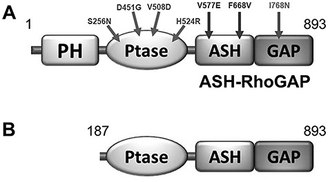
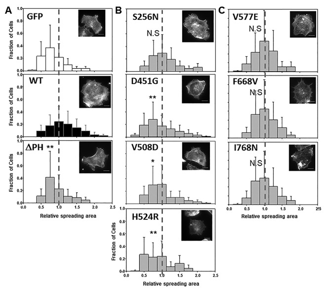
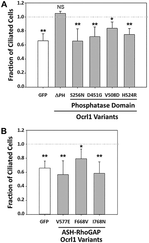
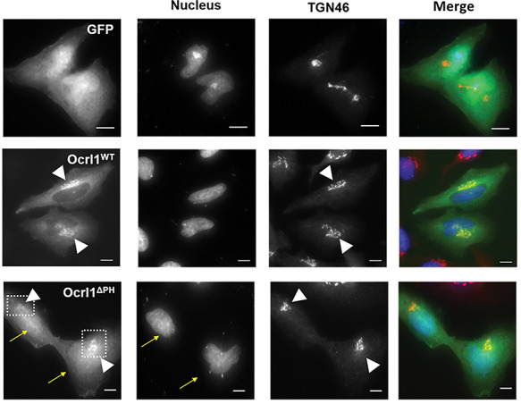
Lowe Syndrome (LS) is a lethal genetic disorder caused by mutations in the OCRL1 gene which encodes the lipid 5' phosphatase Ocrl1. Patients exhibit a characteristic triad of symptoms including eye, brain and kidney abnormalities with renal failure as the most common cause of premature death. Over 200 OCRL1 mutations have been identified in LS, but their specific impact on cellular processes is unknown. Despite observations of heterogeneity in patient symptom severity, there is little understanding of the correlation between genotype and its impact on phenotype. Here, we show that different mutations had diverse effects on protein localization and on triggering LS cellular phenotypes. In addition, some mutations affecting specific domains imparted unique characteristics to the resulting mutated protein. We also propose that certain mutations conformationally affect the 5'-phosphatase domain of the protein, resulting in loss of enzymatic activity and causing common and specific phenotypes (a conformational disease scenario). This study is the first to show the differential effect of patient 5'-phosphatase mutations on cellular phenotypes and introduces a conformational disease component in LS. This work provides a framework that explains symptom heterogeneity and can help stratify patients as well as to produce a more accurate prognosis depending on the nature and location of the mutation within the OCRL1 gene.


Figures

Figure 1
Figure 1
Ocrl1 variants used in this study. A: Residue change due to LS patient missense mutations are mapped on the Ocrl1 molecule. B: Representation of a proposed Ocrl1 variant resulting from alternative initiation at Met187. PH: Pleckstrin homology; Ptase: Inositol phosphatase domain; ASH: ASPM-SPD- 2-Hydin; RhoGAP: RhoGTPase Activating Protein.
Figure 2
Figure 2
Ocrl1 patient variants differentially affect cell spreading. HEK293T KO cells were transfected with Ocrl1WT, Ocrl1ΔPH (A); phosphatase domain mutants (B); or ASH-RhoGAP domain mutants (C) and allowed to attach and spread on fibronectin-coated surfaces (See Materials and Methods). Median spreading areas of cells expressing mutants were normalized with respect to Ocrl1WT. Histograms were constructed using data from three independent experiments, with a total n = 120–150 cells each. Example of rhodamine-phalloidin stained cells representative of the high-frequency groups within each histogram. Scale bar: 10 μm. Statistically significance of the mean difference with respect to Ocrl1WT was A: **P < 0.05 by KS test; B: **P < 0.05, *P < 0.1 (Bonferroni corrected by 4 comparisons), N.S = not significant by KS test; C: N.S = not significant. The vertical reference line highlights WT-normalized median position.
Figure 3
Figure 3
Ocrl1 patient variants differentially affect ciliogenesis. HEK293T KO cells were transfected with different Ocrl1WT, Ocrl1ΔPH and Ocrl1 phosphatase domain mutants (A) or ASH-RhoGAP domain mutants (B). Ciliogenesis assays were performed as described in Materials and Methods. A total of 20 random fields with at least 50 cells were imaged and the fraction of transfected cells with cilia was calculated. This number was normalized to the fraction of Ocrl1WT-expressing cells forming cilia. Each experiment was repeated at least thrice (n = 120–150 cells). Statistically significance of the mean difference with respect to Ocrl1WT was A: **P < 0.05, *P < 0.1 (Bonferroni corrected for 6 comparisons), N.S = not significant by Student’s t-test; B: **P < 0.05, *P < 0.1 (Bonferroni corrected for four comparisons) by the Student’s t-test. The horizontal reference line represents the normalized fraction of Ocrl1WT expressing cells making cilia.
Figure 4
Figure 4
Truncation of Ocrl1 PH domain results in nuclear mislocalization of variant. HK2 KO cells transiently expressing GFP, Ocrl1WT or Ocrl1ΔPH and immunostained for TGN (see Materials and Methods). The arrows indicate Ocrl1ΔPH enrichment in the nuclear compartment; the arrowheads point to TGN and TGN-colocalizing Ocrl1 within the boxed area. Scale bar: 10 μm.
Figure 5
Figure 5
Phosphatase domain mutants produce Golgi apparatus fragmentation. A: HK2 KO cells transiently transfected with GFP-tagged Ocrl1WT, Ocrl1H524R, Ocrl1D451G, Ocrl1V508D or Ocrl1S256N were immunostained for TGN. The highlighted region in merged images corresponds to the TGN area which was enlarged 3X (inset images) to better visualize Golgi apparatus fragmentation. The arrowheads indicate fragmented TGN puncta that lack Ocrl1. The arrows indicate dispersed mutant Ocrl1 lacking TGN colocalization. Scale bar: 10 μm. B-C: The ability of the indicated Ocrl1 patient variants (see horizontal axis at bottom of panel C) to induce GA fragmentation (B) and to catalyze PI(4,5)P2 hydrolysis (C) was tested. B: At least 40 cells transfected with the indicated variants (see horizontal axis at bottom of panel C) were imaged randomly per experiment and the area occupied by TGN was quantified relative to the total cell area (See Materials and Methods). Each experiment was repeated at least thrice with total n = 120–150 cells/group. Statistically significance of the mean difference with respect to Ocrl1WT was **P < 0.05, *P < 0.1 (Bonferroni corrected for four comparisons), NS = No significant by the Wilcoxon test. C: Bacterially expressed and purified GST-Ocrl11–563; WT and the different phosphatase mutants were assayed for enzymatic activity in vitro using the malachite green method as described in Materials and Methods. Statistically significance of the mean difference with respect to Ocrl1WT was **P < 0.05 (Bonferroni corrected for four comparisons), NS = No significant by the t-test. D: HK2 KO cells stably expressing GFP-tagged Ocrl1WT, Ocrl1H524R or Ocrl1D451G (see Materials and Methods) were prepared and immunostained with an anti-TGN46 antibody. Scale bar: 10 μm. E: Golgi apparatus fragmentation determined as a function of total cell area. All cells stably expressing Ocrl1 variants were imaged and TGN area was quantified relative to the total cell area (see Materials and Methods). Statistically significance of the mean difference with respect to Ocrl1WT was *P < 0.1 (Bonferroni corrected for two comparisons) by the Wilcoxon test.
Figure 6
Figure 6
ASH-RhoGAP domain mutants exhibit enrichment or accumulation at the centriole under steady state conditions. A: HK2 KO cells transiently expressing Ocrl1WT or ASH-RhoGAP mutants and maintained in serum-supplemented complete media were immunostained for the centriole marker PC-2 (see Materials and Methods). The arrows indicate Ocrl1 at PC-2 labeled structures. B: Transfected cells were randomly imaged from at least 25 fields containing at least 40 cells. Cells with Ocrl1 colocalization to PC-2 were scored and fraction of cells exhibiting colocalization in a field was determined. Statistically significance of the mean difference with respect to Ocrl1WT was **P < 0.05 (Bonferroni corrected for three comparisons) by the t-test. C: HK2 KO cells transiently expressing Ocrl1V577E or Ocrl1I768N exhibiting protein aggregation. The arrows point to GFP-Ocrl1 colocalization with PC-2. D: HK2 KO cells stably expressing Ocrl1I768N were immunostained for PC-2 (centriole marker) (see Materials and Methods). The highlighted region in merged images corresponds to the perinuclear region which was scaled to 3X (inset images) to better visualize Ocrl1 and PC-2 colocalization. The arrows indicate Ocrl1 at PC-2 labeled structures. Scale bar: 10 μm.
Figure 7
Figure 7
Molecular dynamics prediction of the effect of D451G/V508D mutations on Ocrl1 phosphatase domain structure. A-B: Conformational change in WT (gray) and mutant (green). The residues in catalytic site are represented by sticks, while point mutated variant D451G (A) or V508D (B) is shown by a space-filled model. C-D: Root mean square fluctuation (RMSF), comparison between WT and mutant D451G (C) or V508D (D) indicated that the flexibility of regions within the active site is affected (gray zones). E: Ocrl1 phosphatase domain mutants are impaired for 5′ phosphatase activity. Enzymatic activity was measured as described in Materials and Methods and Figure 5C. Experiments were done at constant enzyme and substrate concentrations while varying the incubation times. All experiments were done in triplicates and repeated at least thrice. Statistically significance of the mean difference with respect to Ocrl1WT at the latest time point was **P < 0.05 (Bonferroni corrected for three comparisons) by student t-test.
Figure 8
Figure 8
Different missense mutations effect on LS phenotypes. Specific mutations have differential effects on general LS phenotypes such as defects on cell spreading (red) and ciliogenesis (green). Different mutations associated with specific amino acid changes (or alternative protein initiation, e.g. ΔPH) at specific Ocrl1 domains are indicated. The Venn diagrams show patient variants inducing only cell spreading defects (ΔPH), only ciliogenesis abnormalities (e.g. ASH-RhoGAP mutated variants) or both (phosphatase mutated proteins, in yellow). Differential phenotype severity is represented by color tone (boxes on the right), the darker the tone the more severe the phenotype, while white indicates the mildest phenotype. Phenotypes associated with changes in specific Ocrl1 domains are indicated. *Patient variant inducing severe (but only) ciliogenesis phenotypes. Note that in the case of GFP-fusions mislocalization to the cytosol leads to GFP-mediated retention in the nucleus.

References

    1. Attree, O., Olivos, I.M., Okabe, I., Bailey, L.C., Nelson, D.L., Lewis, R.A., McInnes, R.R. and Nussbaum, R.L. (1992) The Lowe's oculocerebrorenal syndrome gene encodes a protein highly homologous to inositol polyphosphate-5-phosphatase. Nature, 358, 239–242. - PubMed
    1. Loi, M. (2006) Lowe syndrome. Orphanet J. Rare Dis., 1, 16. - PMC - PubMed
    1. Lowe, C.U., Terrey, M. and MacLachlan, E.A. (1952) Organic-aciduria, decreased renal ammonia production, hydrophthalmos, and mental retardation; a clinical entity. A.M.A. Am. J. Dis. Child., 83, 164–184. - PubMed
    1. Bökenkamp, A. and Ludwig, M. (2016) The oculocerebrorenal syndrome of Lowe: an update. Pediatr. Nephrol., 31, 2201–2212. - PMC - PubMed
    1. Zhang, X., Jefferson, A.B., Auethavekiat, V. and Majerus, P.W. (1995) The protein deficient in Lowe syndrome is a phosphatidylinositol-4,5-bisphosphate 5-phosphatase. Proc. Natl. Acad. Sci. U. S. A., 92, 4853–4856. - PMC - PubMed

Publication types

Substances