Mineralocorticoid Receptor in Smooth Muscle Contributes to Pressure Overload-Induced Heart Failure
- PMID: 33517669
- PMCID: PMC7887087
- DOI: 10.1161/CIRCHEARTFAILURE.120.007279
Mineralocorticoid Receptor in Smooth Muscle Contributes to Pressure Overload-Induced Heart Failure
Abstract
Background: Mineralocorticoid receptor (MR) antagonists decrease heart failure (HF) hospitalization and mortality, but the mechanisms are unknown. Preclinical studies reveal that the benefits on cardiac remodeling and dysfunction are not completely explained by inhibition of MR in cardiomyocytes, fibroblasts, or endothelial cells. The role of MR in smooth muscle cells (SMCs) in HF has never been explored.
Methods: Male mice with inducible deletion of MR from SMCs (SMC-MR-knockout) and their MR-intact littermates were exposed to HF induced by 27-gauge transverse aortic constriction versus sham surgery. HF phenotypes and mechanisms were measured 4 weeks later using cardiac ultrasound, intracardiac pressure measurements, exercise testing, histology, cardiac gene expression, and leukocyte flow cytometry.
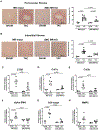
Results: Deletion of MR from SMC attenuated transverse aortic constriction-induced HF with statistically significant improvements in ejection fraction, cardiac stiffness, chamber dimensions, intracardiac pressure, pulmonary edema, and exercise capacity. Mechanistically, SMC-MR-knockout protected from adverse cardiac remodeling as evidenced by decreased cardiomyocyte hypertrophy and fetal gene expression, interstitial and perivascular fibrosis, and inflammatory and fibrotic gene expression. Exposure to pressure overload resulted in a statistically significant decline in cardiac capillary density and coronary flow reserve in MR-intact mice. These vascular parameters were improved in SMC-MR-knockout mice compared with MR-intact littermates exposed to transverse aortic constriction.
Conclusions: These results provide a novel paradigm by which MR inhibition may be beneficial in HF by blocking MR in SMC, thereby improving cardiac blood supply in the setting of pressure overload-induced hypertrophy, which in turn mitigates the adverse cardiac remodeling that contributes to HF progression and symptoms.
Keywords: constriction; endothelial cell; hospitalization; hypertrophy; phenotype.
Figures


Similar articles
-
Aldosterone promotes vascular remodeling by direct effects on smooth muscle cell mineralocorticoid receptors.Arterioscler Thromb Vasc Biol. 2014 Feb;34(2):355-64. doi: 10.1161/ATVBAHA.113.302854. Epub 2013 Dec 5. Arterioscler Thromb Vasc Biol. 2014. PMID: 24311380 Free PMC article.
-
Endothelial mineralocorticoid receptor contributes to systolic dysfunction induced by pressure overload without modulating cardiac hypertrophy or inflammation.Physiol Rep. 2017 Jun;5(12):e13313. doi: 10.14814/phy2.13313. Physiol Rep. 2017. PMID: 28637706 Free PMC article.
-
Smooth Muscle Cell-Mineralocorticoid Receptor as a Mediator of Cardiovascular Stiffness With Aging.Hypertension. 2018 Apr;71(4):609-621. doi: 10.1161/HYPERTENSIONAHA.117.10437. Epub 2018 Feb 20. Hypertension. 2018. PMID: 29463624 Free PMC article.
-
Mineralocorticoid Receptors in Vascular Smooth Muscle: Blood Pressure and Beyond.Hypertension. 2024 May;81(5):1008-1020. doi: 10.1161/HYPERTENSIONAHA.123.21358. Epub 2024 Mar 1. Hypertension. 2024. PMID: 38426347 Free PMC article. Review.
-
Direct role for smooth muscle cell mineralocorticoid receptors in vascular remodeling: novel mechanisms and clinical implications.Curr Hypertens Rep. 2014 May;16(5):427. doi: 10.1007/s11906-014-0427-y. Curr Hypertens Rep. 2014. PMID: 24633842 Free PMC article. Review.
Cited by
-
Capillaries as a Therapeutic Target for Heart Failure.J Atheroscler Thromb. 2022 Jul 1;29(7):971-988. doi: 10.5551/jat.RV17064. Epub 2022 Apr 1. J Atheroscler Thromb. 2022. PMID: 35370224 Free PMC article.
-
Adverse Effects of Aldosterone: Beyond Blood Pressure.J Am Heart Assoc. 2024 Apr 2;13(7):e030142. doi: 10.1161/JAHA.123.030142. Epub 2024 Mar 18. J Am Heart Assoc. 2024. PMID: 38497438 Free PMC article. Review.
-
Multi-omic analysis of the cardiac cellulome defines a vascular contribution to cardiac diastolic dysfunction in obese female mice.Basic Res Cardiol. 2023 Mar 29;118(1):11. doi: 10.1007/s00395-023-00983-6. Basic Res Cardiol. 2023. PMID: 36988733 Free PMC article.
-
Mineralocorticoid receptor blockade normalizes coronary resistance in obese swine independent of functional alterations in Kv channels.Basic Res Cardiol. 2021 May 20;116(1):35. doi: 10.1007/s00395-021-00879-3. Basic Res Cardiol. 2021. PMID: 34018061 Free PMC article.
-
MLK3 mediates impact of PKG1α on cardiac function and controls blood pressure through separate mechanisms.JCI Insight. 2021 Sep 22;6(18):e149075. doi: 10.1172/jci.insight.149075. JCI Insight. 2021. PMID: 34324442 Free PMC article.
References
-
- Pitt B, Zannad F, Remme WJ, Cody R, Castaigne A, Perez A, Palensky J,Wittes J. The effect of spironolactone on morbidity and mortality in patients with severe heart failure. Randomized Aldactone Evaluation Study Investigators. N Engl J Med. 1999;341:709–717. - PubMed
-
- Pitt B, Remme W, Zannad F, Neaton J, Martinez F, Roniker B, Bittman R, Hurley S, Kleiman J, Gatlin M, Eplerenone Post-Acute Myocardial Infarction Heart Failure E,Survival Study I. Eplerenone, a selective aldosterone blocker, in patients with left ventricular dysfunction after myocardial infarction. N Engl J Med. 2003;348:1309–1321. - PubMed
-
- Zannad F, McMurray JJ, Krum H, van Veldhuisen DJ, Swedberg K, Shi H, Vincent J, Pocock SJ, Pitt B,Group E-HS. Eplerenone in patients with systolic heart failure and mild symptoms. N Engl J Med. 2011;364:11–21. - PubMed
-
- Ezekowitz JA,McAlister FA. Aldosterone blockade and left ventricular dysfunction: a systematic review of randomized clinical trials. Eur Heart J. 2009;30:469–477. - PubMed
Publication types
MeSH terms
Substances
Grants and funding
LinkOut - more resources
Full Text Sources
Other Literature Sources
Medical
Research Materials
Miscellaneous

