Observational Study
. 2021 Dec;35(12):3631-3641.
doi: 10.1053/j.jvca.2021.01.011. Epub 2021 Jan 13.

Pulmonary Vascular Thrombosis in COVID-19 Pneumonia

Affiliations
Observational Study

Pulmonary Vascular Thrombosis in COVID-19 Pneumonia

Francesco De Cobelli et al. J Cardiothorac Vasc Anesth. 2021 Dec.

Abstract

Objectives: During severe acute respiratory syndrome coronavirus 2 (SARS-CoV-2) infection, dramatic endothelial cell damage with pulmonary microvascular thrombosis have been was hypothesized to occur. The aim was to assess whether pulmonary vascular thrombosis (PVT) is due to recurrent thromboembolism from peripheral deep vein thrombosis or to local inflammatory endothelial damage, with a superimposed thrombotic late complication.

Design: Observational study.

Setting: Medical and intensive care unit wards of a teaching hospital.

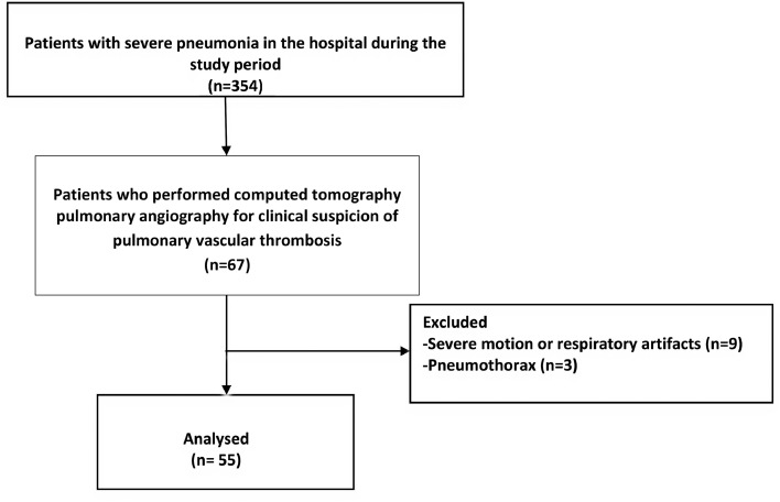
Participants: The authors report a subset of patients included in a prospective institutional study (CovidBiob study) with clinical suspicion of pulmonary vascular thromboembolism.

Interventions: Computed tomography pulmonary angiography and evaluation of laboratory markers and coagulation profile.

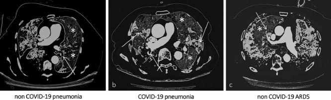
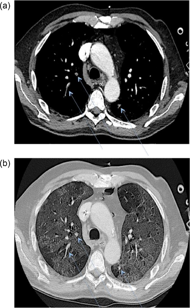
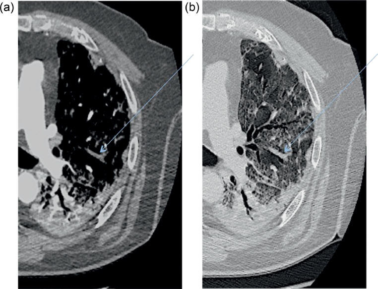
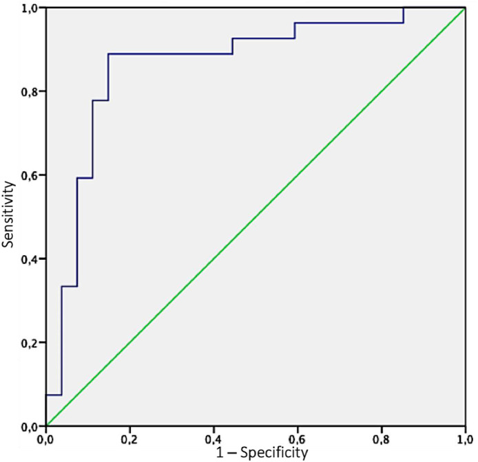
Measurements and main results: Twenty-eight of 55 (50.9%) patients showed PVT, with a median time interval from symptom onset of 17.5 days. Simultaneous multiple PVTs were identified in 22 patients, with bilateral involvement in 16, mostly affecting segmental/subsegmental pulmonary artery branches (67.8% and 96.4%). Patients with PVT had significantly higher ground glass opacity areas (31.7% [22.9-41] v 17.8% [10.8-22.1], p < 0.001) compared with those without PVT. Remarkably, in all 28 patients, ground glass opacities areas and PVT had an almost perfect spatial overlap. D-dimer level at hospital admission was predictive of PVT.

Conclusions: The findings identified a specific radiologic pattern of coronavirus disease 2019 (COVID-19) pneumonia with a unique spatial distribution of PVT overlapping areas of ground-glass opacities. These findings supported the hypothesis of a pathogenetic relationship between COVID-19 lung inflammation and PVT and challenged the previous definition of pulmonary embolism associated with COVID-19 pneumonia.

Keywords: COVID-19; D-dimer increase; computed tomography; critical care; inflammation; thrombosis.


Figures

Image, graphical abstract
Graphical abstract
Fig 1
Fig 1
Flowchart of the study.
Fig 2
Fig 2
Axial contrast-enhanced CT scan demonstrating simultaneous multiple thrombi with bilateral involvement (A) (blue arrows) affecting subsegmental pulmonary artery branches within extensive ground-glass opacities (B).
Fig 3
Fig 3
Axial contrast-enhanced CT scan demonstrating a filling defect (A) (blue arrow) in the left upper lobe overlapping the segmental distribution of ground-glass opacities (B). Notably, no filling defects are present in the left lower lobe, where consolidation is the main radiologic pattern of pneumonia.
Fig 4
Fig 4
The ability of GGOs (ground-glass opacities) ratio in predicting the risk of pulmonary vascular thrombosis was determined by the area under the curve (AUC) of receiver operating characteristics (ROC) curve; the authors identified as best threshold for GGO ratio a percentage of 36.4, above which patients demonstrated a high risk of pulmonary vascular thrombosis (AUC: 0.864; sensitivity: 88.9%; specificity: 85.2%).
Fig 5
Fig 5
Central image/visual abstract: Computed tomography pulmonary angiography (CTPA) imaging demonstrating, in three different clinical scenarios, eventual filling defects in branches of the pulmonary arteries (white arrows) and their spatial distribution according to the corresponding ventilation maps (white asterisks indicate normal lung parenchyma). In (A) (non-COVID-19 pneumonia) and (C) (non-COVID-19 ARDS) filling defects are electively found in the branches of the pulmonary arteries, accountable for the vascularization of healthy lung segments. On the contrary, in (B) (COVID-19 pneumonia), there is an almost perfect topographical overlap between filling defects distribution and pneumonia extent. COVID-19, coronavirus disease 2019.

Similar articles

Cited by

  • The 5 Reasons Why People Die of Coronavirus Disease 2019.
    Marmiere M, D'Amico F, Zangrillo A, Landoni G. Marmiere M, et al. J Cardiothorac Vasc Anesth. 2021 Nov;35(11):3454-3455. doi: 10.1053/j.jvca.2021.03.045. Epub 2021 Apr 1. J Cardiothorac Vasc Anesth. 2021. PMID: 33966995 Free PMC article. No abstract available.
  • Risk of Cardiovascular Events After COVID-19.
    Tereshchenko LG, Bishop A, Fisher-Campbell N, Levene J, Morris CC, Patel H, Beeson E, Blank JA, Bradner JN, Coblens M, Corpron JW, Davison JM, Denny K, Earp MS, Florea S, Freeman H, Fuson O, Guillot FH, Haq KT, Kim M, Kolseth C, Krol O, Lin L, Litwin L, Malik A, Mitchell E, Mohapatra A, Mullen C, Nix CD, Oyeyemi A, Rutlen C, Tam AE, Van Buren I, Wallace J, Khan A. Tereshchenko LG, et al. Am J Cardiol. 2022 Sep 15;179:102-109. doi: 10.1016/j.amjcard.2022.06.023. Epub 2022 Jul 15. Am J Cardiol. 2022. PMID: 35843735 Free PMC article.
  • High Risk of Secondary Infections Following Thrombotic Complications in Patients With COVID-19.
    Ripa M, Galli L, D'Angelo A, Apruzzi L, Palumbo D, Campochiaro C, Tassan Din C, Danise A, Da Prat V, Vitali G, Brugliera L, Poli A, Monardo R, Monti G, Baccellieri D, De Cobelli F, Clementi M, Iannaccone S, Dagna L, Rovere-Querini P, Ciceri F, Tresoldi M, Zangrillo A, Scarpellini P, Castagna A; COVID-BioB Study Group. Ripa M, et al. Open Forum Infect Dis. 2022 Sep 2;9(9):ofac454. doi: 10.1093/ofid/ofac454. eCollection 2022 Sep. Open Forum Infect Dis. 2022. PMID: 36168552 Free PMC article.
  • Anticoagulation as secondary prevention of massive lung thromboses in hospitalized patients with COVID-19.
    Sofia R, Carbone M, Landoni G, Zangrillo A, Dagna L. Sofia R, et al. Eur J Intern Med. 2022 Jun;100:21-24. doi: 10.1016/j.ejim.2022.04.009. Epub 2022 Apr 13. Eur J Intern Med. 2022. PMID: 35487806 Free PMC article. No abstract available.
  • One-Year Multidisciplinary Follow-Up of Patients With COVID-19 Requiring Invasive Mechanical Ventilation.
    Zangrillo A, Belletti A, Palumbo D, Calvi MR, Guzzo F, Fominskiy EV, Ortalda A, Nardelli P, Ripa M, Baiardo Redaelli M, Borghi G, Landoni G, D'Amico F, Marmiere M, Righetti B, Rocchi M, Saracino M, Tresoldi M, Dagna L, De Cobelli F; COVID-BioB Study Group. Zangrillo A, et al. J Cardiothorac Vasc Anesth. 2022 May;36(5):1354-1363. doi: 10.1053/j.jvca.2021.11.032. Epub 2021 Nov 27. J Cardiothorac Vasc Anesth. 2022. PMID: 34973891 Free PMC article.

References

    1. Guan WJ, Ni ZY, Hu Y, et al. Clinical characteristics of coronavirus disease 2019 in China. N Engl J Med. 2020;382:1708–1720. - PMC - PubMed
    1. Ciceri F, Beretta L, Scandroglio AM, et al. Microvascular COVID-19 lung vessels obstructive thromboinflammatory syndrome (MicroCLOTS): An atypical acute respiratory distress syndrome working hypothesis. Crit Care Resusc. 2020;22:95–97. - PMC - PubMed
    1. Varga Z, Flammer AJ, Steiger P, et al. Endothelial cell infection and endotheliitis in COVID-19. Lancet. 2020;395:1417–1418. - PMC - PubMed
    1. Klok FA, Kruip MJHA, Van der Meer NJM, et al. Incidence of thrombotic complications in critically ill ICU patients with COVID-19. Thromb Res. 2020;191:145–147. - PMC - PubMed
    1. Kashi M, Jacquin A, Dakhil B, et al. Severe arterial thrombosis associated with Covid-19 infection. Thromb Res. 2020;192:75–77. - PMC - PubMed

Publication types