

Histologically Proven Dendriform Pulmonary Ossification: A Five-case Series
- PMID: 33518560
- PMCID: PMC8355403
- DOI: 10.2169/internalmedicine.5906-20
Histologically Proven Dendriform Pulmonary Ossification: A Five-case Series
Abstract
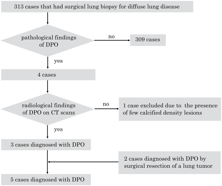
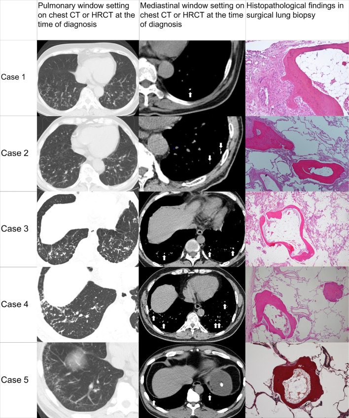
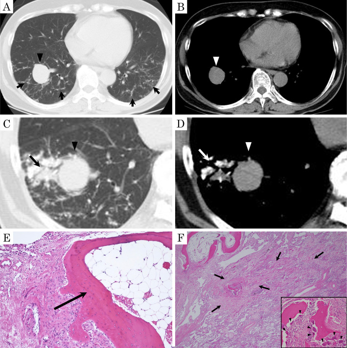
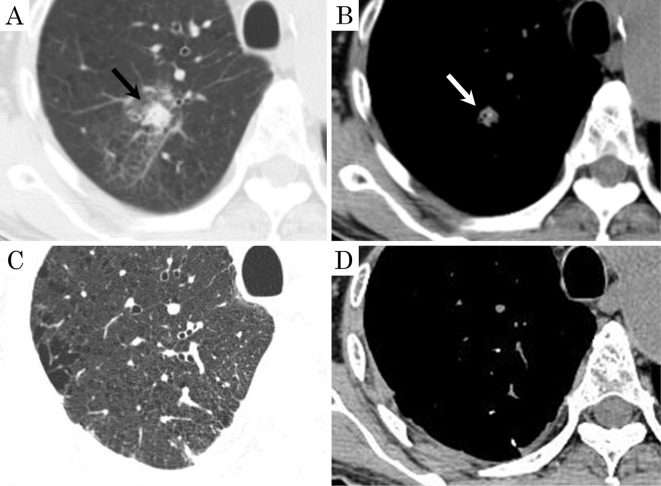
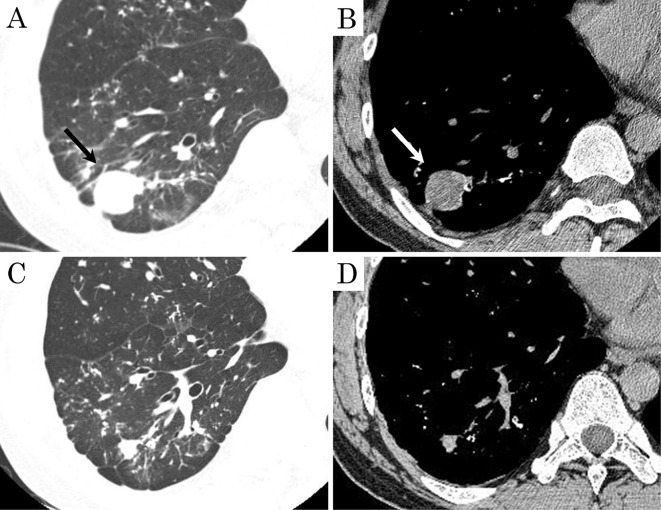
Dendriform pulmonary ossification (DPO) is a rare condition characterized by metaplastic bone formation in the lung parenchyma. It has been reported to be often associated with primary lung diseases, such as usual interstitial pneumonia (UIP) or chronic aspiration of gastric acid; however, its clinical features and pathophysiology remain unclear, especially in idiopathic cases. We herein report five DPO cases, including three with an idiopathic origin. In all cases of idiopathic DPO, the pathological and radiological examinations showed localized pulmonary lesions suggesting inflammation or hemorrhaging.
Keywords: dendriform pulmonary ossification; fibrosis; hemorrhaging; inflammation; surgical lung biopsy; usual interstitial pneumonia.
Conflict of interest statement
Toru Arai: Honoraria, Boehringer Ingelheim and Shionogi. Yoshikazu Inoue: Honoraria, Boehringer Ingelheim, Shionogi and Asahi Kasei.
Figures
References
-
- Lara JF, Catroppo JF, Kim DU, da Costa D. Dendriform pulmonary ossification, a form of diffuse pulmonary ossification: report of a 26-year autopsy experience. Arch Pathol Lab Med 129: 348-353, 2005. - PubMed
-
- Chan ED, Morales DV, Welsh CH, McDermott MT, Schwarz MI. Calcium deposition with or without bone formation in the lung. Am J Respir Crit Care Med 165: 1654-1669, 2002. - PubMed
-
- Gruden JF, Green DB, Legasto AC, Jensen EA, Panse PM. Dendriform pulmonary ossification in the absence of usual interstitial pneumonia: CT features and possible association with recurrent acid aspiration. Am J Roentgenol 209: 1209-1215, 2017. - PubMed
-
- Ndimbie OK, Williams CR, Lee MW. Dendriform pulmonary ossification. Arch Pathol Lab Med 111: 1062-1064, 1987. - PubMed
MeSH terms
LinkOut - more resources
Full Text Sources
Other Literature Sources
Medical