

Hilar Malignant Biliary Obstruction Treated with Four Metallic Stents Involving a New Slim Device
- PMID: 33518571
- PMCID: PMC8263184
- DOI: 10.2169/internalmedicine.6356-20
Hilar Malignant Biliary Obstruction Treated with Four Metallic Stents Involving a New Slim Device
Abstract
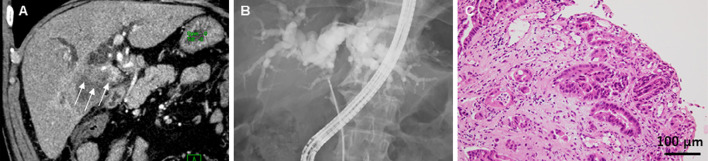
Endoscopic hilar multiple stenting is challenging. A 68-year-old patient had self-expandable metallic stents (SEMSs) inserted for unresectable hilar malignant biliary obstruction. After the SEMSs were inserted into the left hepatic duct and bile duct branch of segment (B) 6, a new SEMS with a wide mesh and slim delivery system was inserted into the right anterior hepatic duct. However, liver abscess and dilated B7 were observed on computed tomography; therefore, an additional new SEMS was quickly and easily inserted into B7. After the placement of these four SEMSs, the liver abscess improved. The new SEMS was effective for hilar multiple biliary drainage.
Keywords: bilateral biliary drainage; hilar malignant biliary obstruction; self-expandable metallic stent.
Conflict of interest statement
Figures


Similar articles
-
Predictive factors for the failure of endoscopic stent-in-stent self-expandable metallic stent placement to treat malignant hilar biliary obstruction.World J Gastroenterol. 2017 Sep 14;23(34):6273-6280. doi: 10.3748/wjg.v23.i34.6273. World J Gastroenterol. 2017. PMID: 28974893 Free PMC article.
-
Direct comparison of simultaneous and sequential endoscopic metallic bilateral stenting in malignant hilar biliary obstruction.World J Gastroenterol. 2025 May 21;31(19):101913. doi: 10.3748/wjg.v31.i19.101913. World J Gastroenterol. 2025. PMID: 40497097 Free PMC article.
-
Endoscopic Stenting of a Fully Covered Self-Expandable Metal Stent with a Hole in Each Cavity in Malignant Hilar Biliary Obstruction: A Preclinical Proof-of-Concept Study and Initial Human Experience.Dig Dis Sci. 2025 Mar;70(3):1215-1222. doi: 10.1007/s10620-024-08810-1. Epub 2025 Jan 24. Dig Dis Sci. 2025. PMID: 39856481
-
Endoscopic biliary stenting for unresectable malignant hilar obstruction.Clin J Gastroenterol. 2017 Dec;10(6):485-490. doi: 10.1007/s12328-017-0778-4. Epub 2017 Oct 19. Clin J Gastroenterol. 2017. PMID: 29052124 Review.
-
Topic controversies in the endoscopic management of malignant hilar strictures using metal stent: side-by-side versus stent-in-stent techniques.J Hepatobiliary Pancreat Sci. 2015 Sep;22(9):650-6. doi: 10.1002/jhbp.270. Epub 2015 Jul 1. J Hepatobiliary Pancreat Sci. 2015. PMID: 26136361 Review.
Cited by
-
Efficacy of a novel large-cell Niti-S stent with a slim delivery system for hilar biliary obstruction: a preliminary study.Ann Med. 2022 Dec;54(1):953-961. doi: 10.1080/07853890.2022.2056631. Ann Med. 2022. PMID: 35412414 Free PMC article.
References
-
- Nuzzo G, Giuliante F, Ardito F, et al. . Improvement in perioperative and long-term outcome after surgical treatment of hilar cholangiocarcinoma: results of an Italian multicenter analysis of 440 patients. Arch Surg 147: 26-34, 2012. - PubMed
-
- Takahashi Y, Nagino M, Nishio H, Ebata T, Igami T, Nimura Y. Percutaneous transhepatic biliary drainage catheter tract recurrence in cholangiocarcinoma. Br J Surg 97: 1860-1866, 2010. - PubMed
-
- Kawakami H, Kuwatani M, Onodera M, et al. . Endoscopic nasobiliary drainage is the most suitable preoperative biliary drainage method in the management of patients with hilar cholangiocarcinoma. J Gastroenterol 46: 242-248, 2011. - PubMed
Publication types
MeSH terms
LinkOut - more resources
Full Text Sources
Other Literature Sources
Medical
Research Materials