

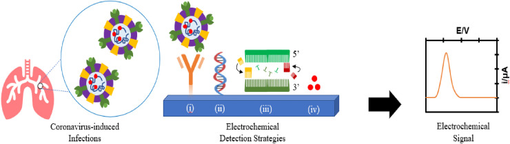
The potential of electrochemistry for the detection of coronavirus-induced infections
- PMID: 33518851
- PMCID: PMC7836945
- DOI: 10.1016/j.trac.2020.116081
The potential of electrochemistry for the detection of coronavirus-induced infections
Abstract
Human coronaviruses (HCoV) are no stranger to the global environment. The etiology of previous outbreaks with reported symptoms of respiratory tract infections was attributed to different coronavirus strains, with the latest global pandemic in 2019 also belonging to the coronavirus family. Timely detection, effective therapeutics and future prevention are stake key holders in the management of coronavirus-induced infections. Apart from the gold standard clinical diagnostics, electrochemical techniques have also demonstrated their great potentials in the detection of different viruses and their correlated antibodies and antigens, showing high sensitivities and selectivities, and faster times for the analysis. This article aims to critically review the multifaceted electrochemical approaches, not only in the development of point-of-care portable devices but also as alternative detection strategies that can be coupled with traditional methods for the detection of various strains of coronaviruses.
Keywords: Biosensing; Biosensors; COVID-19; Coronavirus; Detection; Diagnostic; Electrochemical methods; Electrochemistry; Infections; SARS-CoV-2.
© 2020 Elsevier B.V. All rights reserved.
Conflict of interest statement
The authors declare no conflict of interest.
Figures
Similar articles
-
Current Strategies of Antiviral Drug Discovery for COVID-19.Front Mol Biosci. 2021 May 13;8:671263. doi: 10.3389/fmolb.2021.671263. eCollection 2021. Front Mol Biosci. 2021. PMID: 34055887 Free PMC article. Review.
-
Point-of-Care Diagnostics of COVID-19: From Current Work to Future Perspectives.Sensors (Basel). 2020 Jul 31;20(15):4289. doi: 10.3390/s20154289. Sensors (Basel). 2020. PMID: 32752043 Free PMC article. Review.
-
Laboratory detection methods for the human coronaviruses.Eur J Clin Microbiol Infect Dis. 2021 Feb;40(2):225-246. doi: 10.1007/s10096-020-04001-8. Epub 2020 Sep 28. Eur J Clin Microbiol Infect Dis. 2021. PMID: 32984911 Free PMC article. Review.
-
Current and Future Point-of-Care Tests for Emerging and New Respiratory Viruses and Future Perspectives.Front Cell Infect Microbiol. 2020 Apr 29;10:181. doi: 10.3389/fcimb.2020.00181. eCollection 2020. Front Cell Infect Microbiol. 2020. PMID: 32411619 Free PMC article. Review.
-
Etiology and clinical characteristics of SARS-CoV-2 and other human coronaviruses among children in Zhejiang Province, China 2017-2019.Virol J. 2021 Apr 30;18(1):89. doi: 10.1186/s12985-021-01562-8. Virol J. 2021. PMID: 33931105 Free PMC article.
Cited by
-
A Short Review Comparing Carbon-Based Electrochemical Platforms With Other Materials For Biosensing SARS-Cov-2.ChemistrySelect. 2022 Oct 7;7(37):e202202465. doi: 10.1002/slct.202202465. Epub 2022 Oct 4. ChemistrySelect. 2022. PMID: 36711230 Free PMC article. Review.
-
Nanobiocide Based-Silver Nanomaterials Upon Coronaviruses: Approaches for Preventing Viral Infections.Nanoscale Res Lett. 2021 Jun 6;16(1):100. doi: 10.1186/s11671-021-03558-3. Nanoscale Res Lett. 2021. PMID: 34095961 Free PMC article. Review.
-
Biosensors as Nano-Analytical Tools for COVID-19 Detection.Sensors (Basel). 2021 Nov 24;21(23):7823. doi: 10.3390/s21237823. Sensors (Basel). 2021. PMID: 34883826 Free PMC article. Review.
-
A review on the recent achievements on coronaviruses recognition using electrochemical detection methods.Microchem J. 2022 Jul;178:107322. doi: 10.1016/j.microc.2022.107322. Epub 2022 Feb 25. Microchem J. 2022. PMID: 35233118 Free PMC article. Review.
-
Paper-based analytical devices for virus detection: Recent strategies for current and future pandemics.Trends Analyt Chem. 2021 Nov;144:116424. doi: 10.1016/j.trac.2021.116424. Epub 2021 Aug 26. Trends Analyt Chem. 2021. PMID: 34462612 Free PMC article. Review.
References
-
- Liu D.X., Liang J.Q., Fung T.S. Elsevier; 2020. Human Coronavirus-229E, -OC43, -NL63, and -HKU1.
-
- Lu R., Zhao X., Li J., Niu P., Yang B., Wu H., Wang W., Song H., Huang B., Zhu N., Bi Y., Ma X., Zhan F., Wang L., Hu T., Zhou H., Hu Z., Zhou W., Zhao L., Chen J., Meng Y., Wang J., Lin Y., Yuan J., Xie Z., Ma J., Liu W.J., Wang D., Xu W., Holmes E.C., Gao G.F., Wu G., Chen W., Shi W., Tan W. Genomic characterisation and epidemiology of 2019 novel coronavirus: implications for virus origins and receptor binding. Lancet. 2020;395:565–574. - PMC - PubMed
-
- Rockx B., Kuiken T., Herfst S., Bestebroer T., Lamers M.M., Oude Munnink B.B., de Meulder D., van Amerongen G., van den Brand J., Okba N.M.A., Schipper D., van Run P., Leijten L., Sikkema R., Verschoor E., Verstrepen B., Bogers W., Langermans J., Drosten C., Fentener van Vlissingen M., Fouchier R., de Swart R., Koopmans M., Haagmans B.L. Comparative pathogenesis of COVID-19, MERS, and SARS in a nonhuman primate model. Science. 2020;368:1012–1015. - PMC - PubMed
-
- Zhou P., Yang X.L., Wang X.G., Hu B., Zhang L., Zhang W., Si H.R., Zhu Y., Li B., Huang C.L., Chen H.D., Chen J., Luo Y., Guo H., Jiang R.D., Liu M.Q., Chen Y., Shen X.R., Wang X., Zheng X.S., Zhao K., Chen Q.J., Deng F., Liu L.L., Yan B., Zhan F.X., Wang Y.Y., Xiao G.F., Shi Z.L. A pneumonia outbreak associated with a new coronavirus of probable bat origin. Nature. 2020;579:270–273. - PMC - PubMed
Publication types
LinkOut - more resources
Full Text Sources
Miscellaneous