. 2021 Jan 15:14:623134.
doi: 10.3389/fncel.2020.623134. eCollection 2020.

Antidromic Spike Propagation and Dissimilar Expression of P2X, 5-HT, and TRPV1 Channels in Peripheral vs. Central Sensory Axons in Meninges

Affiliations

Antidromic Spike Propagation and Dissimilar Expression of P2X, 5-HT, and TRPV1 Channels in Peripheral vs. Central Sensory Axons in Meninges

Oleg Gafurov et al. Front Cell Neurosci. .

Abstract

Background: The terminal branches of the trigeminal nerve in meninges are supposed to be the origin site of migraine pain. The main function of these peripheral sensory axons is the initiation and propagation of spikes in the orthodromic direction to the second order neurons in the brainstem. The stimulation of the trigeminal ganglion induces the release of the neuropeptide CGRP in meninges suggesting the antidromic propagation of excitation in these fibers. However, the direct evidence on antidromic spike traveling in meningeal afferents is missing. Methods: By recording of spikes from peripheral or central parts of the trigeminal nerve in rat meninges, we explored their functional activity and tested the expression of ATP-, serotonin-, and capsaicin-gated receptors in the distal vs. proximal parts of these nerves. Results: We show the significant antidromic propagation of spontaneous spikes in meningeal nerves which was, however, less intense than the orthodromic nociceptive traffic due to higher number of active fibers in the latter. Application of ATP, serotonin and capsaicin induced a high frequency nociceptive firing in peripheral processes while, in central parts, only ATP and capsaicin were effective. Disconnection of nerve from trigeminal ganglion dramatically reduced the tonic antidromic activity and attenuated the excitatory action of ATP. Conclusion: Our data indicate the bidirectional nociceptive traffic and dissimilar expression of P2X, 5-HT and TRPV1 receptors in proximal vs. distal parts of meningeal afferents, which is important for understanding the peripheral mechanisms of migraine pain.

Keywords: 5-HT; ATP; TRPV1 receptor; excitability; meninges; migraine; trigeminal nerve.


Conflict of interest statement

The authors declare that the research was conducted in the absence of any commercial or financial relationships that could be construed as a potential conflict of interest.

Figures

Figure 1
Figure 1
Orthodromic and antidromic propagation of action potentials in the trigeminal nerve. (A) Original images of the rat meninges with the middle meningeal artery (MMA), trigeminal ganglion (TG), and N. spinosus peripheral (a) or central parts (b) of the trigeminal nerve placed inside the recording electrode. Right, schematic presentation of the functional destination of the orthodromic and antidromic spike propagation. (B) Example traces of spontaneous action potentials propagated in the trigeminal nerve either in the orthodromic (a) and antidromic (b) directions. (C) Histograms showing comparison of the baseline frequency of orthodromic (Ortho, n = 10, white column) and antidromic action potentials (Anti, n = 17, black column) directions in the trigeminal nerve (**p < 0.01). (D) Histograms showing comparison of the baseline frequency of orthodromic (n = 10, white column) and antidromic action potentials (n = 17, black column) in the trigeminal nerve; mean ± SEM, **p < 0.01, Mann-Whitney test.
Figure 2
Figure 2
Testing the action of ATP and serotonin (5-HT) in peripheral (orthodromic) and central (antidromic) parts of trigeminal afferents. (A) Example traces of orthodromic action potentials in the trigeminal nerve in control (a), after application 100 μM ATP (b) and 2 μM 5-HT (c). (B) Example traces of antidromic action potentials in the trigeminal nerve in control (a), after application of 100 μM ATP (b) and 2 μM 5-HT (c). (C) The frequency of orthodromic (n = 10, black cycles) and antidromic (n = 17, white cycle) action potentials during application of 100 μM ATP. (D) The frequency of orthodromic (n = 10, black cycles) and antidromic (n = 10, white cycles) action potentials during application of 2 μM 5-HT; mean ± SEM, *p < 0.05, **p < 0.01, t-test.
Figure 3
Figure 3
Cluster analysis of spiking activity in the trigeminal nerve in the orthodromic and antidromic directions. (A) The example distribution of clusters (plot of the amplitude of the negative spike phase vs. positive phase) in the orthodromic (a) and antidromic (b) firing. Notice a smaller number of clusters during antidromic firing. (B) Histograms showing comparison of the total number of clusters in the orthodromic (Ortho, n = 10, white column) firing and antidromic (Anti, n = 17, black column) firing; mean ± SEM, **p < 0.01, Mann-Whitney test.
Figure 4
Figure 4
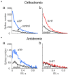
Spectral analysis of ATP and serotonin (5-HT) induced orthodromic and antidromic firing. (A) The distribution of interspike intervals (ISI) for orthodromic firing in control and after application of 100 μM ATP (a; n = 10) and 2 μM 5-HT (b; n = 10). (B) The distribution of interspike intervals (ISI) for antidromic activity in control and after 100 μM ATP [n = 17 (a) and for μM 5-HT (b, n = 10)]. Notice lack of changes in spectral distribution of antidromic spikes for 5-HT.

References

    1. Ault B., Evans R. H. (1980). Depolarizing action of capsaicin on isolated dorsal-root fibers of the rat. J. Physiol. 306, 22–23.
    1. Basbaum A. I., Woolf C. J. (1999). Pain. Curr Biol. 9, 29–31. 10.1016/S0960-9822(99)80273-5 - DOI - PubMed
    1. Bernardini N., Neuhuber W., Reeh P. W., Sauer S. K. (2004). Morphological evidence for functional capsaicin receptor expression and calcitonin gene-related peptide exocytosis in isolated peripheral nerve axons of the mouse. Neuroscience 126, 585–590. 10.1016/j.neuroscience.2004.03.017 - DOI - PubMed
    1. Cinar E., Zhou S., DeCourcey J., Wang Y., Waugh R. E., Wan J. (2015). Piezo1 regulates mechanotransductive release of ATP from human RBCs. Proc. Natl. Acad. Sci. U.S.A. 112, 11783–11788. 10.1073/pnas.1507309112 - DOI - PMC - PubMed
    1. Della Pietra A., Mikhailov N., Giniatullin R. (2020). The emerging role of mechanosensitive piezo channels in migraine pain. Int. J. Mol. Sci. 21:696. 10.3390/ijms21030696 - DOI - PMC - PubMed