. 2021 Jan 13:11:594479.
doi: 10.3389/fphar.2020.594479. eCollection 2020.

Hemokinin-1 as a Mediator of Arthritis-Related Pain via Direct Activation of Primary Sensory Neurons

Affiliations

Hemokinin-1 as a Mediator of Arthritis-Related Pain via Direct Activation of Primary Sensory Neurons

Éva Borbély et al. Front Pharmacol. .

Abstract

The tachykinin hemokinin-1 (HK-1) is involved in immune cell development and inflammation, but little is known about its function in pain. It acts through the NK1 tachykinin receptor, but several effects are mediated by a yet unidentified target. Therefore, we investigated the role and mechanism of action of HK-1 in arthritis models of distinct mechanisms with special emphasis on pain. Arthritis was induced by i.p. K/BxN serum (passive transfer of inflammatory cytokines, autoantibodies), intra-articular mast cell tryptase or Complete Freund's Adjuvant (CFA, active immunization) in wild type, HK-1- and NK1-deficient mice. Mechanical- and heat hyperalgesia determined by dynamic plantar esthesiometry and increasing temperature hot plate, respectively, swelling measured by plethysmometry or micrometry were significantly reduced in HK-1-deleted, but not NK1-deficient mice in all models. K/BxN serum-induced histopathological changes (day 14) were also decreased, but early myeloperoxidase activity detected by luminescent in vivo imaging increased in HK-1-deleted mice similarly to the CFA model. However, vasodilation and plasma protein extravasation determined by laser Speckle and fluorescent imaging, respectively, were not altered by HK-1 deficiency in any models. HK-1 induced Ca2+-influx in primary sensory neurons, which was also seen in NK1-deficient cells and after pertussis toxin-pretreatment, but not in extracellular Ca2+-free medium. These are the first results showing that HK-1 mediates arthritic pain and cellular, but not vascular inflammatory mechanisms, independently of NK1 activation. HK-1 activates primary sensory neurons presumably via Ca2+ channel-linked receptor. Identifying its target opens new directions to understand joint pain leading to novel therapeutic opportunities.

Keywords: arthritic pain; experimental arthritis; in vivo optical imaging; neuroinflammation; primary sensory neuron; tachykinin.


Conflict of interest statement

Zsuzsanna Helyes is the strategic director and shareholder of PharmInVivo Ltd. (Pécs, Hungary) and shareholder of Algonist Biotechnologies Gmbh, (Wien, Austria). Eva Szoke is also a shareholder of Algonist Biotechnologies Gmbh, (Wien, Austria), but there is no conflict of interest with the present work. These companies were not involved in the study design, funding, collection, analysis, interpretation of data, the writing of this article or the decision to submit it for publication. The remaining authors declare that the research was conducted in the absence of any commercial or financial relationships that could be construed as a potential conflict of interest.

Figures
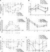
FIGURE 1
FIGURE 1
Change in mechanonociceptive threshold (A), heat threshold (B), paw volume (C), cold tolerance (D), body weight (E) and time spent on grid (F) in K/BxN serum-induced arthritis of wild type (WT) and hemokinin-1-deficient (Tac4 −/−) mice in comparison with BxN serum-treated non-arthritic groups (n = 4–10 per group; *p < 0.05, **p < 0.01, ***p < 0.001 vs. arthritic WT, repeated measures two-way ANOVA + Bonferroni’s post test).
FIGURE 2
FIGURE 2
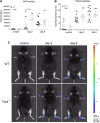
Quantitative evaluation of MPO-activity (A) and IR-676 dye extravasation (B) in K/BxN arthritogenic serum-treated wild type (WT) and hemokinin-1-deficient (Tac4 −/−) mice. Representative images of MPO-activity (C). Box plots demonstrate medians with the upper and lower quartiles and all individual data points; n = 6–12 per group; *p < 0.05 vs. WT, ##p < 0.01, ###p < 0.001 vs. control (one-way ANOVA + Bonferroni’s post test).
FIGURE 3
FIGURE 3
Representative histopathological pictures of safranin O-stained tibiotarsal joint showing ×100 and ×200 magnifications with 500 and 200 µm scale bars, respectively. ti = tibia, ta = tarsus, s = synovium (A). Semiquantitative histopathological score of K/BxN serum treated wild type (WT) and hemokinin-deficient (Tac4 −/−) arthritic mice in comparison with non-arthritic controls. Box plots demonstrate the medians with upper and lower quartiles of n = three to five per group (*p < 0.05 vs. WT, Mann-Whitney test) (B).
FIGURE 4
FIGURE 4
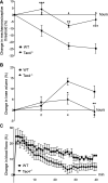
Changes in mechanonociceptive threshold (A), knee diameter (B) and blood flow (C) in mast cell tryptase-induced acute arthritis of wild type (WT) and hemokinin-1-deficient (Tac4 −/−) mice. Data points demonstrate the means ± SEM of n = 5–10 per group; *p < 0.05, **p < 0.01, ***p < 0.001 vs. WT (repeated measures two-way ANOVA + Bonferroni’s post test).
FIGURE 5
FIGURE 5
Changes in mechanonociceptive threshold (A), AP knee diameter (B), and MPO-activity (C) in CFA-induced acute arthritis of WT and HK-1-deficient (Tac4 −/−) mice. Data points demonstrate the means ± SEM (A), (B) or medians with upper and lower quartiles of n = 8–15 per group; *p < 0.05, **p < 0.01, ***p < 0.001 vs. WT, ###p < 0.001 vs. control (repeated measures two-way ANOVA + Bonferroni’s post test; for MPO activity: one-way ANOVA + Bonferroni’s post test).
FIGURE 6
FIGURE 6
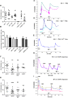
Effect of HK-1 and SP in cultured primary sensory neurons. Change in the fluorescence ratio (R = F340/F380) in HK-1-sensitive cells (•) is presented. ●: HK-1 and PTX co-administration, ♦: fluorescence signal in neurons from NK1R −/− animals, ○: HK-1 and CP99994 NK1 receptor antagonist administration, □: HK-1 and AMG9810 TRPV1 receptor antagonist, ■: HK-1 and HC 030031 TRPA1 receptor antagonist. No signal was detected in Ca2+-free extracellular solution. N = 19–49 cells/group (A). The percentage of responsive cells to HK-1, HK-1+PTX, HK-1 in Ca2+-free condition, HK-1+NK1 receptor antagonist CP99994 and HK-1 in TG from NK1R −/− animals, HK-1 + TRPV1 receptor antagonist AMG9810 and HK-1 + TRPA1 receptor antagonist HC 030031 is presented. Ca2+-responses are presented in % of total number of examined neurons. N = 19–49 cells per group (B). Original Ca2+-imaging registrations after HK-1 administration. Increases of R = 340/380 fluorescence in fura-2 loaded cultured TG neurons (C). Original Ca2+-imaging registrations after HK-1 administration in TG neurons from NK1 gene deficient mice (D). Original Ca2+-imaging registrations after HK-1 administration in Ca2+-free solution (E). Effect of HK-1 on capsaicin-induced Ca2+-influx. Increases of R = 340/380 fluorescence in fura-2 loaded neurons are presented, **p < 0.01, NS (vs. caps1, one-way ANOVA with Bonferroni’s multiple comparison post hoc test, n = 16) (F). Effect of HK-1 on capsaicin-induced Ca2+-influx. Original Ca2+-imaging registrations after capsaicin and HK-1 administration (G). Effect of SP on capsaicin-induced Ca2+-influx. Increases of R = 340/380 fluorescence in fura-2 loaded neurons are presented, ***p < 0.001, NS (vs. caps1, one-way ANOVA with Bonferroni’s multiple comparison post hoc test, n = 17) (H). Effect of SP on capsaicin-induced Ca2+-influx. Original Ca2+-imaging registrations after capsaicin and SP administration (I).

References

    1. Aczél T., Kecskés A., Kun J., Szenthe K., Bánáti F., Szathmary S., et al. (2020). Hemokinin-1 gene expression is upregulated in trigeminal ganglia in an inflammatory orofacial pain model: potential role in peripheral sensitization. Int. J. Mol. Sci. 21 (8), 2938 10.3390/ijms21082938 - DOI - PMC - PubMed
    1. Agalave N. M., Larsson M., Abdelmoaty S., Su J., Baharpoor A., Lundbäck P., et al. (2014). Spinal HMGB1 induces TLR4-mediated long-lasting hypersensitivity and glial activation and regulates pain-like behavior in experimental arthritis. Pain 155, 1802–1813. 10.1016/j.pain.2014.06.007 - DOI - PubMed
    1. Arnhold J., Flemmig J. (2010). Human myeloperoxidase in innate and acquired immunity. Arch. Biochem. Biophys. 500, 92–106. 10.1016/j.abb.2010.04.008 - DOI - PubMed
    1. Berger A., Benveniste P., Corfe S. A., Tran A. H., Barbara M., Wakeham A., et al. (2010). Targeted deletion of the tachykinin 4 gene (TAC4-/-) influences the early stages of B lymphocyte development. Blood 116, 3792–3801. 10.1182/blood-2010-06-291062 - DOI - PubMed
    1. Berger A., Tran A. H., Paige C. J. (2007). Co-regulated decrease of Neurokinin-1 receptor and Hemokinin-1 gene expression in monocytes and macrophages after activation with pro-inflammatory cytokines. J. Neuroimmunol. 187, 83–93. 10.1016/j.jneuroim.2007.04.019 - DOI - PubMed