. 2021 Jan 14:11:606592.
doi: 10.3389/fneur.2020.606592. eCollection 2020.

Atypical Resting-State Functional Connectivity Dynamics Correlate With Early Cognitive Dysfunction in HIV Infection

Affiliations

Atypical Resting-State Functional Connectivity Dynamics Correlate With Early Cognitive Dysfunction in HIV Infection

Benedictor Alexander Nguchu et al. Front Neurol. .

Abstract

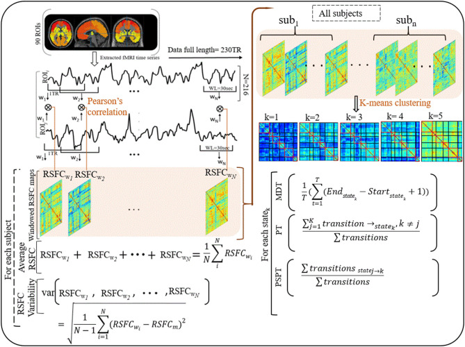
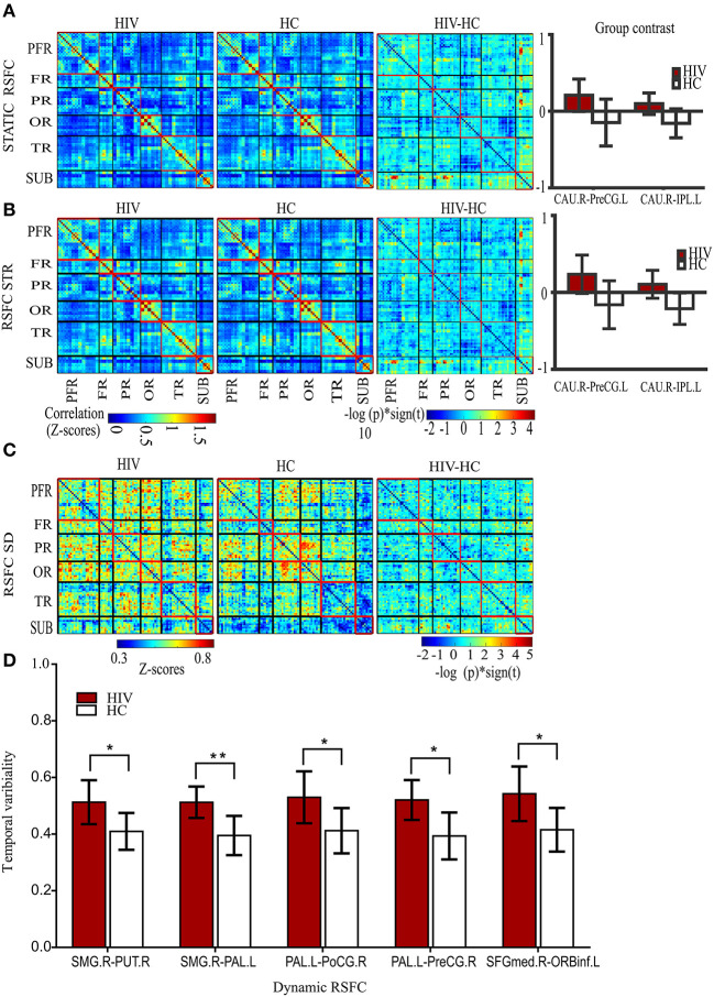
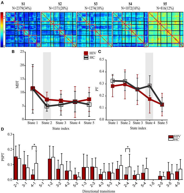
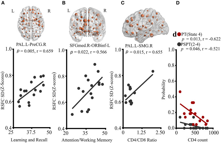
Purpose: Previous studies have shown that HIV affects striato-cortical regions, leading to persisting cognitive impairment in 30-70% of the infected individuals despite combination antiretroviral therapy. This study aimed to investigate brain functional dynamics whose deficits might link to early cognitive decline or immunologic deterioration. Methods: We applied sliding windows and K-means clustering to fMRI data (HIV patients with asymptomatic neurocognitive impairment and controls) to construct dynamic resting-state functional connectivity (RSFC) maps and identify states of their reoccurrences. The average and variability of dynamic RSFC, and the dwelling time and state transitioning of each state were evaluated. Results: HIV patients demonstrated greater variability in RSFC between the left pallidum and regions of right pre-central and post-central gyri, and between the right supramarginal gyrus and regions of the right putamen and left pallidum. Greater variability was also found in the frontal RSFC of pars orbitalis of the left inferior frontal gyrus and right superior frontal gyrus (medial). While deficits in learning and memory recall of HIV patients related to greater striato-sensorimotor variability, deficits in attention and working memory were associated with greater frontal variability. Greater striato-parietal variability presented a strong link with immunologic function (CD4+/CD8+ ratio). Furthermore, HIV-infected patients exhibited longer time and reduced transitioning in states typified by weaker connectivity in specific networks. CD4+T-cell counts of the HIV-patients were related to reduced state transitioning. Conclusion: Our findings suggest that HIV alters brain functional connectivity dynamics, which may underlie early cognitive impairment. These findings provide novel insights into our understanding of HIV pathology, complementing the existing knowledge.

Keywords: HIV-associated neurocognitive disorders; brain resting-state functional connectivity dynamics; fMRI; functional connectivity; temporal variability.


Conflict of interest statement

The authors declare that the research was conducted in the absence of any commercial or financial relationships that could be construed as a potential conflict of interest.

Figures

Figure 1
Figure 1
Illustrations of procedures for dynamic RSFC and state analyses. Ninty ROIs' times series were extracted from fMRI data (230 TR) and sliding windows (WL = 30 s) were used to generate dynamic RSFC maps. Dynamic RSFC properties (average dynamic RSFC, and connectivity variability) were computed across windows. Optimal clusters (states, k = 1–5) were generated using K-means clustering. State properties [the mean dwelling time of the state (MDT), and state transitioning (PT, PSPT)] were further estimated from each state. RSFCwi represents dynamic RSFC map at window wi; MDT and PT denote the mean dwelling and probability of transitioning to statek respectively, whereas PSPT signifies the probability of specific transitioning from statej to statek.
Figure 2
Figure 2
Static and dynamic functional connectivity. (A) The static RSFC maps for HIV and HC groups, group difference map (HIV-HC), and between-group contrast. (B) The average dynamic RSFC maps for HIV and HC groups, group difference maps (HIV-HC), and between-group contrast in average dynamic RSFC. (C) The mean dynamic temporal variability maps for HIV and HC groups, and group variability-difference maps (HIV-HC). (D) The group difference for temporal variability as depicted in the histogram. PFR, prefrontal regions; FR, other frontal regions; PR, parietal regions; OR, occipital regions; TR, parietal regions; and SUB, subcortical regions. R, right; L, left; CAU, the caudate nucleus; IPL, inferior frontal lobe; and PreCG, precentral gyrus; SMG, supramarginal gyrus, PUT, putamen; PAL, pallidum; PoCG, postcentral gyrus; SFGmed; superior frontal (medial) gyrus; ORBinf, the pars of orbitalis of the inferior frontal gyrus. Pearson's correlation coefficients were all fisher's z-transformed. The group-difference (HIV-HC) connectivity maps are displayed as –log10 (p-value) × sign (t).
Figure 3
Figure 3
States properties of HIV infected individuals and healthy controls. (A) Five transient states generated by K-means clustering analysis, displayed with the number of correlation maps constituting to the state. (B) Mean dwelling time (MDT) between groups. (C) The probabilities of transitioning (PT) for each state in HIV and HC groups. (D) The probabilities of specific state-to-state transitioning (PSPT). On top of the figures are the numbers (N=) of dynamic correlation maps [with their percentages (%)] showing the population of correlation maps assigned to those states during clustering. Group differences (HIV-HC) were significant at p < 0.05.
Figure 4
Figure 4
Correlations of properties of dynamic RSFC and RSFC states with clinical characteristics of HIV patients. (A) Correlation of PAL.L-to-PreCG.R temporal variability with learning and recall score. (B) Association of temporal variability of SFGmed.R-to-ORBinf-L with Attention/working memory performance. (C) The relationship between PAL.L-SMG.R variability and CD4+/CD8+ ratio. (D) Relationships of probabilities of transitioning to state 4, and state-2-to-state-4 transitioning with CD4+ T-cell counts. The displayed relationships were evaluated in Pearson's correlations coefficients, r; p < 0.05. P, p-value; L, left; R, right; PAL, the pallidum; PreCG, precentral gyrus; SFGmed, superior frontal (medial) gyrus; ORBinf, pars orbitalis of the inferior frontal gyrus; and SMG, supramarginal gyrus.

Similar articles

Cited by

References

    1. Abidin AZ, Dsouza AM, Nagarajan MB, Wang L, Qiu X, Schifitto G, et al. . Alteration of brain network topology in HIV-associated neurocognitive disorder: a novel functional connectivity perspective. Neuroimage Clin. (2018) 17:768–777. 10.1016/j.nicl.2017.11.025 - DOI - PMC - PubMed
    1. Antinori A, Arendt G, Becker JT, Brew BJ, Byrd DA, Cherner M, et al. . Updated research nosology for HIV-associated neurocognitive disorders. Neurology. (2007) 69:1789–99. 10.1212/01.WNL.0000287431.88658.8b - DOI - PMC - PubMed
    1. Wiesman AI, O'Neill J, Mills MS, Robertson KR, Fox HS, Swindells S, et al. . Aberrant occipital dynamics differentiate HIV-infected patients with and without cognitive impairment. Brain. (2018) 141:1678–90. 10.1093/brain/awy097 - DOI - PMC - PubMed
    1. Ellis R, Langford D, Masliah E. HIV and antiretroviral therapy in the brain: neuronal injury and repair. Nat Rev Neurosci. (2007) 8:33–44. 10.1038/nrn2040 - DOI - PubMed
    1. McIntosh RC, Rosselli M, Uddin LQ, Antoni M. Neuropathological sequelae of human immunodeficiency virus and apathy: a review of neuropsychological and neuroimaging studies. Neurosci Biobehav Rev. (2015) 55:147–64. 10.1016/j.neubiorev.2015.04.008 - DOI - PubMed