Review
. 2021 Jan 14:11:604048.
doi: 10.3389/fmicb.2020.604048. eCollection 2020.

A Place for Viruses on the Tree of Life

Affiliations
Review

A Place for Viruses on the Tree of Life

Hugh M B Harris et al. Front Microbiol. .

Abstract

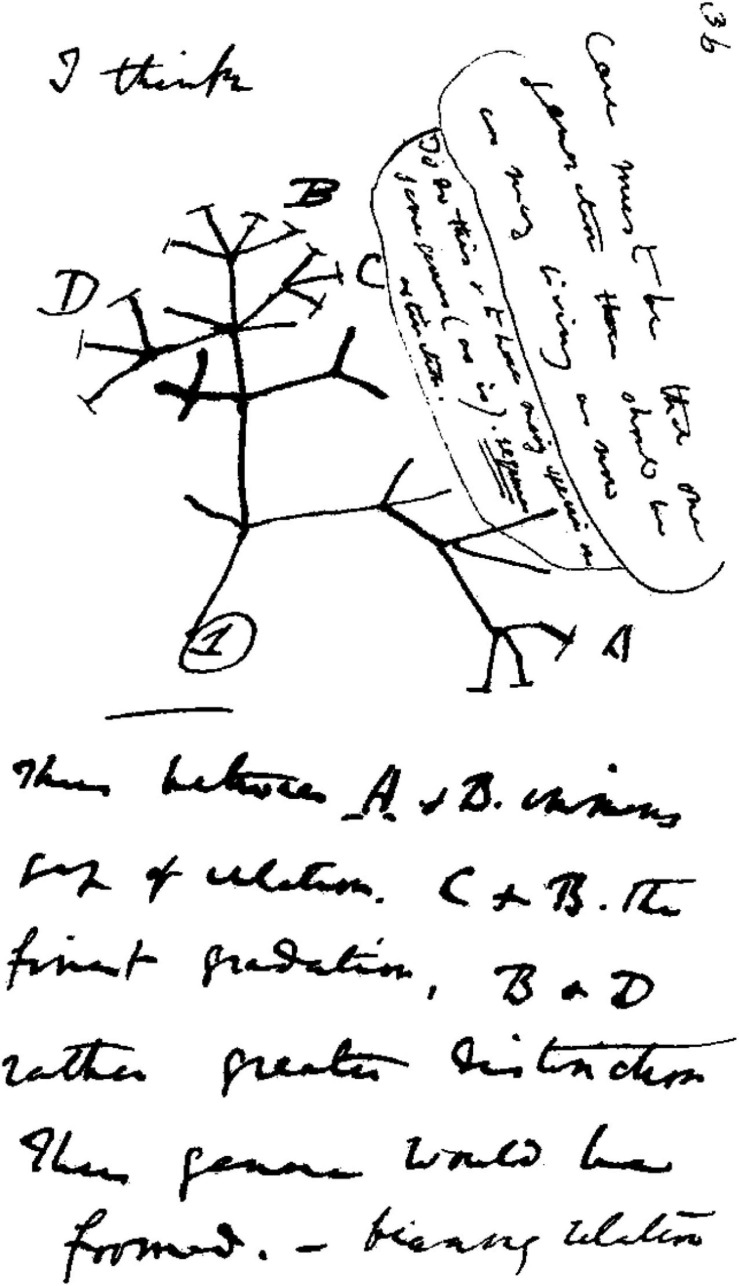
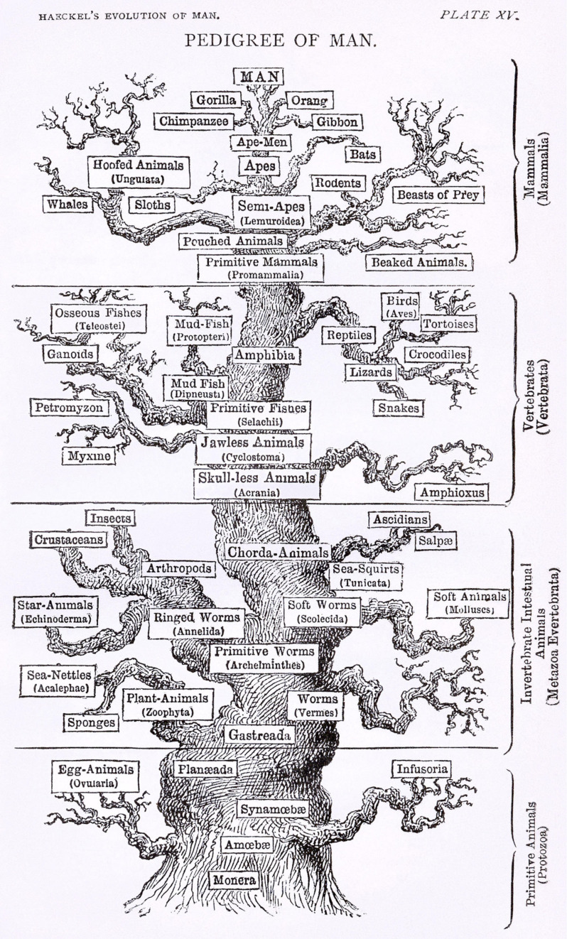
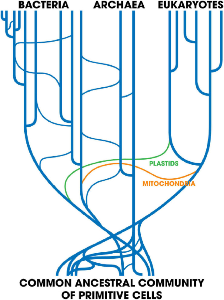
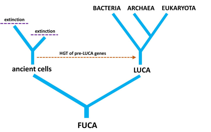
Viruses are ubiquitous. They infect almost every species and are probably the most abundant biological entities on the planet, yet they are excluded from the Tree of Life (ToL). However, there can be no doubt that viruses play a significant role in evolution, the force that facilitates all life on Earth. Conceptually, viruses are regarded by many as non-living entities that hijack living cells in order to propagate. A strict separation between living and non-living entities places viruses far from the ToL, but this may be theoretically unsound. Advances in sequencing technology and comparative genomics have expanded our understanding of the evolutionary relationships between viruses and cellular organisms. Genomic and metagenomic data have revealed that co-evolution between viral and cellular genomes involves frequent horizontal gene transfer and the occasional co-option of novel functions over evolutionary time. From the giant, ameba-infecting marine viruses to the tiny Porcine circovirus harboring only two genes, viruses and their cellular hosts are ecologically and evolutionarily intertwined. When deciding how, if, and where viruses should be placed on the ToL, we should remember that the Tree functions best as a model of biological evolution on Earth, and it is important that models themselves evolve with our increasing understanding of biological systems.

Keywords: Tree of Life; evolution; horizontal gene transfer; phylogeny; viruses.


Conflict of interest statement

The authors declare that the research was conducted in the absence of any commercial or financial relationships that could be construed as a potential conflict of interest.

Figures

FIGURE 1
FIGURE 1
Charles Darwin’s 1837 sketch. His first diagram of an evolutionary tree from his First Notebook on Transmutation of Species (1837). Interpretation of handwriting: “I think case must be that one generation should have as many living as now. To do this and to have as many species in same genus (as is) requires extinction. Thus between A + B the immense gap of relation. C + B the finest gradation. B + D rather greater distinction. Thus genera would be formed.” Attribution: Charles Darwin/Public domain.
FIGURE 2
FIGURE 2
A version of the “tree of life” by Ernst Haeckel, from The Evolution of Man (1879). Attribution: Ernst Haeckel/Public domain.
FIGURE 3
FIGURE 3
Taken from Smets and Barkay (2005). Horizontal gene transfer: Perspectives at a crossroads of scientific disciplines. Created: 23 May 2018; License: CC-BY-SA 4.0.
FIGURE 4
FIGURE 4
A simple tree diagram showing the relationship between LUCA (last universal common ancestor) and FUCA (first universal cellular ancestor). The hypothetical transfer of pre-LUCA genes from ancient cells into post-LUCA genomes is also depicted, along with extinction events of cellular lineages that have no modern descendants.
FIGURE 5
FIGURE 5
Taken from Iranzo et al. (2016). Internal structure of the Caudovirales supermodule. A bipartite graph is displayed, linking bacterial modules to the genes they share. Proteins that represent hub nodes in the gene-sharing network are labeled. License: CC Attribution 4.0 International License.
FIGURE 6
FIGURE 6
A simple tree diagram showing the chimeric origin of viruses from pre-LUCA replication genes and post-LUCA structural genes. Ancient MGE ancestors replace ancient cells (from Figure 4), reflecting the origin of virus replication genes from MGEs. The evolution of modern plasmids and transposons from ancient MGEs is also depicted.
FIGURE 7
FIGURE 7
A simple diagram of the phylogenetic forest of gene trees with network components linking different genes together. The diagram shows four gene lineages, two viral and two cellular. Arrows connect genes present on the same genome to their respective viral or bacterial species, while the lysogenic virus is connected to species A, showing that it is a prophage whose genes reside on the same genome as species A.

References

    1. Arinkin V., Smyshlyaev G., Barabas O. (2019). Jump ahead with a twist: DNA acrobatics drive transposition forward. Curr. Opin. Struct. Biol. 59 168–177. 10.1016/j.sbi.2019.08.006 - DOI - PMC - PubMed
    1. Bapteste E., Susko E., Leigh J., Macleod D., Charlebois R. L., Doolittle W. F. (2005). Do orthologous gene phylogenies really support tree-thinking? BMC Evol. Biol. 5:33. 10.1186/1471-2148-5-33 - DOI - PMC - PubMed
    1. Benner S. A. (2010). Defining life. Astrobiology 10 1021–1030. - PMC - PubMed
    1. Berche P. (2012). Louis Pasteur, from crystals of life to vaccination. Clin. Microbiol. Infect. 18(Suppl. 5) 1–6. 10.1111/j.1469-0691.2012.03945.x - DOI - PubMed
    1. Berkemer S. J., McGlynn S. E. (2020). A new analysis of archaea-bacteria domain separation: variable phylogenetic distance and the tempo of early evolution. Mol. Biol. Evol. 37 2332–2340. 10.1093/molbev/msaa089 - DOI - PMC - PubMed