. 2021 Jan 15:11:611823.
doi: 10.3389/fgene.2020.611823. eCollection 2020.

LncRNA H19 Suppresses Osteosarcomagenesis by Regulating snoRNAs and DNA Repair Protein Complexes

Affiliations

LncRNA H19 Suppresses Osteosarcomagenesis by Regulating snoRNAs and DNA Repair Protein Complexes

An Xu et al. Front Genet. .

Abstract

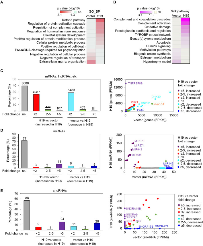
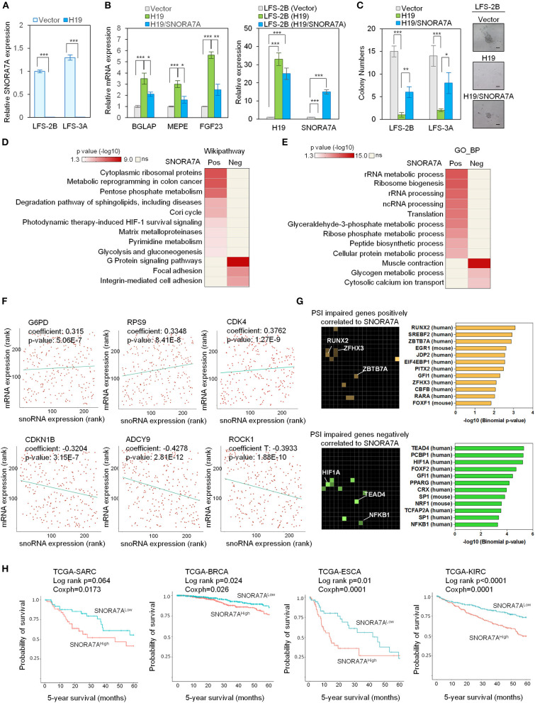
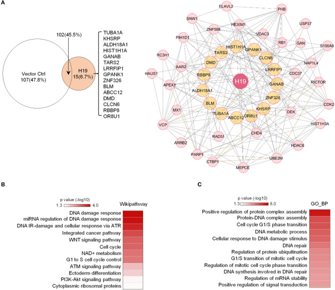
Osteosarcoma is one of the most frequent common primary malignant tumors in childhood and adolescence. Long non-coding RNAs (lncRNAs) have been reported to regulate the initiation and progression of tumors. However, the exact molecular mechanisms involving lncRNA in osteosarcomagenesis remain largely unknown. Li-Fraumeni syndrome (LFS) is a familial cancer syndrome caused by germline p53 mutation. We investigated the tumor suppressor function of lncRNA H19 in LFS-associated osteosarcoma. Analyzing H19-induced transcriptome alterations in LFS induced pluripotent stem cell (iPSC)-derived osteoblasts, we unexpectedly discovered a large group of snoRNAs whose expression was significantly affected by H19. We identified SNORA7A among the H19-suppressed snoRNAs. SNORA7A restoration impairs H19-mediated osteogenesis and tumor suppression, indicating an oncogenic role of SNORA7A. TCGA analysis indicated that SNORA7A expression is associated with activation of oncogenic signaling and poor survival in cancer patients. Using an optimized streptavidin-binding RNA aptamer designed from H19 lncRNA, we revealed that H19-tethered protein complexes include proteins critical for DNA damage response and repair, confirming H19's tumor suppressor role. In summary, our findings demonstrate a critical role of H19-modulated SNORA7A expression in LFS-associated osteosarcomas.

Keywords: H19 lncRNA; Li-Fraumeni syndrome; iPSCs; osteosarcoma; p53; snoRNA.


Conflict of interest statement

The authors declare that the research was conducted in the absence of any commercial or financial relationships that could be construed as a potential conflict of interest.

Figures

Figure 1
Figure 1
H19 significantly affects miRNA and snoRNA expression. (A,B) Heatmaps show the GO_BP (A) and Wikipathway (B) categories enriched in either H19-expressing LFS osteoblasts (right column) or vector control LFS osteoblasts (left column) compared with each other based on mRNA expression of respective samples. Colors represent p values of enrichment. (C-E) H19 overexpression induces small changes in overall mRNA expression but large changes in miRNA and snoRNA expression in LFS osteoblasts. The percentage of mRNAs, lncRNAs, etc. (C), miRNAs (D), and snoRNAs (E) with different fold-changes (<2, 2-5, and >5 increase or decrease) are shown in bar plots. RNA expression between H19-overexpressing and vector control cells is presented in a scatter plot (right). Dot colors are used to represent the fold change of RNA expression. ns (not significant) indicates mRNAs, lncRNAs, miRNAs, snoRNAs, etc. with fold-change <2 and/or FPKM<1. The RNA-seq experiment was done with one sample pooled from 3 biological replicates. miRNAs and snoRNAs are defined based on the miRbase and snoDB database, respectively. “mRNA, lncRNA, etc.” indicates all transcripts except miRNAs and snoRNAs.
Figure 2
Figure 2
Systems analyses of the oncogenic role of SNORA7A from TCGA datasets. (A) Ectopic expression of H19 downregulates SNORA7A expression in LFS osteoblasts. qRT-PCR data are represented as mean ±SEM; n = 3 biological replicates; statistical significance is determined using two-tailed Student's t-test; ***p < 0.001. (B) Left panel, ectopic expression of H19 increases osteogenic gene expression in LFS osteoblasts; in contrast, restoration of SNORA7A expression impairs H19-upregulated osteogenic gene expression in H19-transduced LFS osteoblasts. Right panel, qRT-PCR results demonstrate the expression of H19 and SNORA7A upon their ectopic expression for assays in the left panel. qRT-PCR data are represented as mean ± SEM; n = 3 biological replicates; statistical significance is determined using two-tailed Student's t-test; *p < 0.05; **p < 0.01; ***p < 0.001. (C) AIG assay for in vitro tumorigenicity demonstrates that H19 impairs the colony numbers of LFS osteoblasts and SNORA7A expression rescues the H19-suppressed tumorigenicity of LFS osteoblasts. H19 or H19/SNORA7A-transduced LFS osteoblasts were grown for 1 month and then assayed. Positive colonies are considered those larger than 50μm diameter. Data are represented as mean ± SEM; n = 6 biological replicates; statistical significance is determined using two-tailed Student's t-test; *p < 0.05; **p < 0.01; ***p < 0.001. (D,E) Genes positively (n = 780) and negatively (n = 945) correlated with SNORA7A expression in TCGA-SARC are identified. Pathway analysis by Wikipathway (A) and GO_BP (B) is performed on these gene sets using Enrichr to identify pathways significantly enriched or depleted (p ≤ 0.05) in association with SNORA7A expression. (F) Scatterplots of G6PD, RPS9, CDK4, CDKN1B, ADCY9, and ROCK1 mRNA expression correlation with SNORA7A expression. (G) Genes whose abnormal splicing (including exon skip, mutually exclusive splicing, or intron retention) by PSI was positively (n = 91, upper panel) or negatively (n = 182, lower panel) correlated with SNORA7A expression in sarcomas are identified by TRANSFAC and JASPAR PWWMs in Enrichr. PSI: percent spliced in index. (H) High SNORA7A expression is associated with poor cancer survival. Five-year overall survival is analyzed according to SNORA7A expression from TCGA in various cancers. SARC: sarcoma. BRCA: breast invasive carcinoma. ESCA: esophageal carcinoma. KIRC: kidney renal clear cell carcinoma.
Figure 3
Figure 3
Potential H19 interacting proteins are primarily involved in DNA damage/repair response. (A) Left panel: Mass spectrometry analysis of TetO-FUW-4S1m-H19 streptavidin pull-down proteins identifies 15 unique H19-interacting proteins. TetO-FUW-4S1m vector is used as an S1m pull-down negative control (left). Right panel: The biological protein-protein interaction network based on H19-interacting proteins (orange, n = 15) and expanded to include genes (pink, n = 31) connected to at least two H19-interacting proteins is constructed in Cytoscape using the Biogrid human database. A solid line indicates physical interactions. A dashed line indicates genetic interactions. (B,C) Heatmaps representing p-values of enriched genesets (p ≤ 0.05) among the H19-interacting protein complex genes (n = 47) by Wikipathway (B) and GO_BP (C) analyses.

References

    1. Amelio I., Mancini M., Petrova V., Cairns R. A., Vikhreva P., Nicolai S., et al. . (2018). p53 mutants cooperate with HIF-1 in transcriptional regulation of extracellular matrix components to promote tumor progression. Proc. Natl. Acad. Sci. U.S.A 115, E10869–E10878. 10.1073/pnas.1808314115 - DOI - PMC - PubMed
    1. Bachellerie J. P., Cavaille J., Huttenhofer A. (2002). The expanding snoRNA world. Biochimie 84, 775–790. 10.1016/s0300-9084(02)01402-5 - DOI - PubMed
    1. Carvajal-Vergara X., Sevilla A., D'Souza S. L., Ang Y. S., Schaniel C., Lee D. F., et al. . (2010). Patient-specific induced pluripotent stem-cell-derived models of LEOPARD syndrome. Nature 465, 808–812. 10.1038/nature09005 - DOI - PMC - PubMed
    1. Chen E. Y., Tan C. M., Kou Y., Duan Q., Wang Z., Meirelles G. V., et al. . (2013). Enrichr: interactive and collaborative HTML5 gene list enrichment analysis tool. BMC Bioinform. 14:128. 10.1186/1471-2105-14-128 - DOI - PMC - PubMed
    1. Chen P. L., Liu F., Cai S., Lin X., Li A., Chen Y., et al. . (2005). Inactivation of CtIP leads to early embryonic lethality mediated by G1 restraint and to tumorigenesis by haploid insufficiency. Mol. Cell Biol. 25, 3535–3542. 10.1128/MCB.25.9.3535-3542.2005 - DOI - PMC - PubMed