

doi: 10.18502/jovr.v16i1.8263.
eCollection 2021 Jan-Mar.
An Unusual Presentation of Vogt-Koyanagi-Harada
Affiliations
- PMID: 33520140
- PMCID: PMC7841286
- DOI: 10.18502/jovr.v16i1.8263
Item in Clipboard
An Unusual Presentation of Vogt-Koyanagi-Harada
J Ophthalmic Vis Res.
.
No abstract available
Conflict of interest statement
There are no conflicts of interest.
Figures
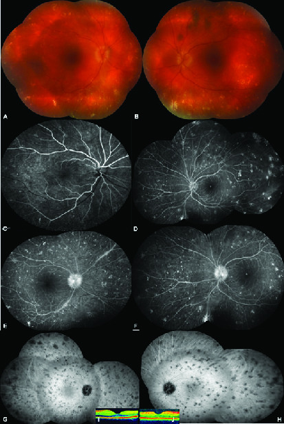
Composite of color fundus showing 360° scattered yellowish–gray spot like aggregates, (A) right eye, (B) left eye. Early venous phase of composite fluorescein angiographic picture exhibiting staining of these spot-like lesions, (C) right eye, (D) left eye. Late venous phase of composite fluorescein angiographic picture showing leakage from the optic disc, (E) (right eye), (F) left eye. Mid-phase of composite indocyanine green picture demonstrating the hypocyanescent widespread spot-like opacities, (G) right eye, (H) left eye. Normal foveal contour on optical coherence tomography, (I) right eye, (J) left eye.
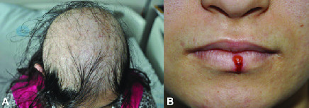
(A) Appearance of hair scalp from above depicting the diffuse alopecia. (B) Colored picture of coexistent Herpes Labialis.
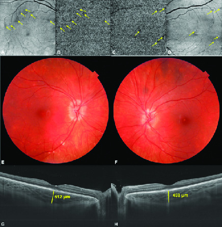
(A&B) Choriocapillaris slab of montage optical coherence tomography angiography and en-face OCT image of the right. (C&D) Left eyes revealing the patchy ischemia of choriocapillaris (arrows). Color fundus pictures taken at the last visit showing slightly blurred disc margins with less apparent old scars with no new lesion. (E) Right eye and (F) left eye. (G) Swept source optical coherence tomographic subfoveal choroidal thickness was 512 μm in the right eye and (H) 498 μm in the left eye.
References
-
- Read RW, Holland GN, Rao NA, Tabbara KF, Ohno S, Arellanes-Garcia L, et al. Revised diagnostic criteria for Vogt–Koyanagi–Harada disease: report of an international committee on nomenclature. Am J Ophthalmol 2001;131:647–652. - PubMed
-
- DiPreta EA, Smith KJ, Williams J, Skelton H. Histopathologic findings in the alopecia associated with Vogt-Koyanagi-Harada disease. J Cutan Med Surg 2000;4:155–159. - PubMed
-
- Yang P, Zhong Y, Du L, Chi W, Chen L, Zhang R, et al. Development and evaluation of diagnostic criteria
LinkOut - more resources
Full Text Sources
Other Literature Sources