. 2020 Dec 3:9:1392.
doi: 10.12688/f1000research.27504.2. eCollection 2020.

Isolation and primary culture of Galleria mellonella hemocytes for infection studies

Affiliations

Isolation and primary culture of Galleria mellonella hemocytes for infection studies

Nicola J Senior et al. F1000Res. .

Abstract

Galleria mellonella larvae are increasingly used to study the mechanisms of virulence of microbial pathogens and to assess the efficacy of antimicrobials. The G. mellonella model can faithfully reproduce many aspects of microbial disease which are seen in mammals, and therefore allows a reduction in the use of mammals. The model is now being widely used by researchers in universities, research institutes and industry. An attraction of the model is the interaction between pathogen and host. Hemocytes are specialised phagocytic cells which resemble neutrophils in mammals and play a major role in the response of the larvae to infection. However, the detailed interactions of hemocytes with pathogens is poorly understood, and is complicated by the presence of different sub-populations of cells. We report here a method for the isolation of hemocytes from Galleria mellonella. A needle-stick injury of larvae, before harvesting, markedly increased the recovery of hemocytes in the hemolymph. The majority of the hemocytes recovered were granulocyte-like cells. The hemocytes survived for at least 7 days in culture at either 28°C or 37°C. Pre-treatment of larvae with antibiotics did not enhance the survival of the cultured hemocytes. Our studies highlight the importance of including sham injected, rather than un-injected, controls when the G. mellonella model is used to test antimicrobial compounds. Our method will now allow investigations of the interactions of microbial pathogens with insect hemocytes enhancing the value of G. mellonella as an alternative model to replace the use of mammals, and for studies on hemocyte biology.

Keywords: 3Rs; Galleria mellonella; hemocytes; infection model.


Conflict of interest statement

Competing interests: Richard W Titball is a shareholder in Biosystems Technology Ltd

Figures

Figure 1.
Figure 1.. View of a single G. mellonella larva from the underside, showing the six frontal prolegs (arrowed) which are the preferred sites for injection.
Figure 2.
Figure 2.. Technique for immobilisation of larvae for the injection or withdrawal of fluids.
Figure 3.
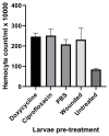
Figure 3.. Hemocytes recovered from larvae pre-dosed with the antibiotic indicated, PBS or wounded with a syringe needle for three days prior to hemocyte recovery.
The results shown are the average of four counts made on hemolymph extracted from groups of 10 larvae per treatment. Error bars = SD.
Figure 4.
Figure 4.
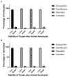
Viability of hemocytes incubated at 28ºC at T0 ( A) or T7 ( B). Average across three replicate wells per treatment, derived from the pooled hemolymph from 10 larvae, and seeded at 2 × 10 5 cells/ml. Hemocytes dissociated with trypsin and stained with trypan blue. Error bars = SD. Mean percentage (and SD) of hemocytes live at T0: doxycycline treated, 93% (±3.2%); ciprofloxacin treated, 98% (±2.5%); wounded, 96% (±3.5%); untreated, 93% (±3.6%). Mean percentage (and SD) of hemocytes live at T7: doxycycline treated, 95% (±4.7%); ciprofloxacin treated, 95% (±2.5%); wounded, 96% (±2.1%); wounded, 94% (±3.5%).
Figure 5.
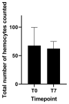
Figure 5.. Total hemocyte count of cells in 10 µl from sham injected larvae incubated at 28°C at T0 and T7.
Average across three replicate wells, derived from the pooled hemolymph from 10 larvae and seeded at 2 × 10 5 cells/ml. Hemocytes stained with trypan blue and dissociated with trypsin at T7 only. Errors bars = SD. Mean (and SD) cell counts of hemocytes at T0; 68 (±32) or at T7; 62 (±12.5).
Figure 6.
Figure 6.
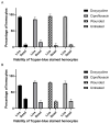
Viability of ex vivo hemocytes incubated at 37°C at T0 ( A) and T7 ( B). Average across 3 replicate wells per treatment, derived from the pooled hemolymph from 10 larvae and seeded at 2 × 10 5 cells/ml. Hemocytes were dissociated with trypsin and stained with trypan blue. Error bars = SD. Mean percentage (and SD) of hemocytes live at T0: doxycycline treated 93% (±3); ciprofloxacin treated, 85% (±6.9); wounded, 95% (±6.2); untreated, 93% (±2.1). Mean percentage (and SD) of hemocytes live at T7: doxycycline treated 71% (±2.5); ciprofloxacin treated, 81% (±4.2); wounded, 79% (±4.4); untreated, 77% (±6.7).

References

    1. Champion OL, Wagley S, Titball RW: Galleria mellonella as a model host for microbiological and toxin research. Virulence. 2016;7(7):840–5. 10.1080/21505594.2016.1203486 - DOI - PMC - PubMed
    1. Tsai CJY, Loh JMS, Proft T: Galleria mellonella infection models for the study of bacterial diseases and for antimicrobial drug testing. Virulence. 2016;7(3):214–29. 10.1080/21505594.2015.1135289 - DOI - PMC - PubMed
    1. Cutuli MA, Petronio GP, Vergalito XX, et al. : Galleria mellonella as a consolidated in vivo model hosts: New developments in antibacterial strategies and novel drug testing. Virulence. 2019;10(1):527–541. 10.1080/21505594.2019.1621649 - DOI - PMC - PubMed
    1. Kavanagh K, Sheehan G: The Use of Galleria mellonella Larvae to Identify Novel Antimicrobial Agents against Fungal Species of Medical Interest. J Fungi (Basel). 2018;4(3):113. 10.3390/jof4030113 - DOI - PMC - PubMed
    1. Allegra E, Titball RW, Carter J, et al. : Galleria mellonella larvae allow the discrimination of toxic and non-toxic chemicals. Chemosphere. 2018;198:469–472. 10.1016/j.chemosphere.2018.01.175 - DOI - PubMed

Publication types

LinkOut - more resources