

Biventricular Outflow Obstruction Associated With Atrioventricular Septal Defects and Patent Ductus Arteriosus: An Extremely Rare Combination
- PMID: 33520483
- PMCID: PMC7834562
- DOI: 10.7759/cureus.12265
Biventricular Outflow Obstruction Associated With Atrioventricular Septal Defects and Patent Ductus Arteriosus: An Extremely Rare Combination
Abstract
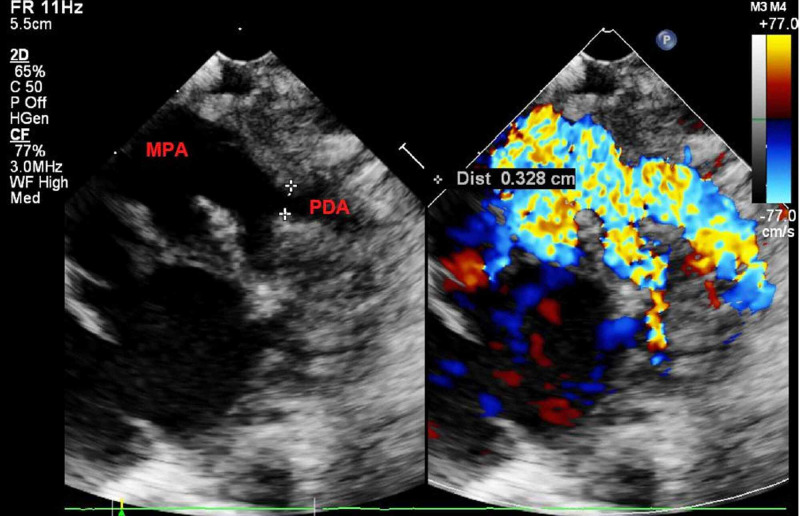
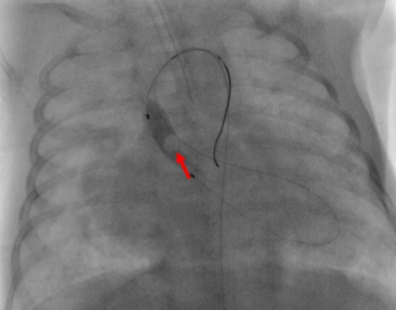
We present an extremely rare combination of biventricular outflow obstruction associated with atrioventricular septal defects and patent ductus arteriosus (PDA). Almost all the other published cases, including ours, were associated with other congenital cardiac lesions other than biventricular outflow obstruction. Most cases ended with poor outcomes. Our patient was a 55-day-old term female infant. She was managed by successful aortic balloon valvuloplasty with successful early outcome.
Keywords: atrioventricular septal defects; balloon valvuloplasty; biventricular outflow obstruction; combined semilunar valves stenosis; congenital heart diseases (chds); neonates; patent ductus arteriosus; pediatric cardiology.
Copyright © 2020, Alabdulgader et al.
Conflict of interest statement
The authors have declared that no competing interests exist.
Figures


References
-
- 20-year survival of children born with congenital anomalies: a population-based study. Tennant PW, Pearce MS, Bythell M, Rankin J. https://pubmed.ncbi.nlm.nih.gov/20092884/ Lancet. 2010;30:649–656. - PubMed
-
- Congenital heart disease in 740 subjects: epidemiological aspects. Alabdulgader AAA. https://pubmed.ncbi.nlm.nih.gov/11471253/ Ann Trop Paediatr. 2001;3:111–118. - PubMed
-
- Abstract 17714: prevalence of bicuspid aortic valve in 9000 newborns and associated echocardiographic findings estimated by systematic echocardiographic screening. Sillesen A-S, Pihl C-A, Vejlstrup NG. https://www.ahajournals.org/doi/10.1161/circ.136.suppl_1.17714 Circulation. 2017;136:0.
-
- Coexisting congenital stenoses of aortic and pulmonic ostia. Richter GW. https://pubmed.ncbi.nlm.nih.gov/13091586/ AMA Arch Pathol. 1953;56:392–396. - PubMed
-
- Congenital valvular stenosis of pulmonary and aortic valves with atrial septal defect. Horlick L, Merriman JE. Am Heart J. 1957;54:615–620. - PubMed
Publication types
LinkOut - more resources
Full Text Sources