. 2021 Jan 15:8:619377.
doi: 10.3389/fchem.2020.619377. eCollection 2020.

Lanthanide-Doped Upconversion Nanoparticles for Super-Resolution Microscopy

Affiliations

Lanthanide-Doped Upconversion Nanoparticles for Super-Resolution Microscopy

Hao Dong et al. Front Chem. .

Abstract

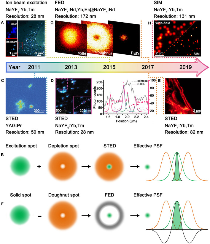
Super-resolution microscopy offers a non-invasive and real-time tool for probing the subcellular structures and activities on nanometer precision. Exploring adequate luminescent probes is a great concern for acquiring higher-resolution image. Benefiting from the atomic-like transitions among real energy levels, lanthanide-doped upconversion nanoparticles are featured by unique optical properties including excellent photostability, large anti-Stokes shifts, multicolor narrowband emissions, tunable emission lifetimes, etc. The past few years have witnessed the development of upconversion nanoparticles as probes for super-resolution imaging studies. To date, the optimal resolution reached 28 nm (λ/36) for single nanoparticles and 82 nm (λ/12) for cytoskeleton structures with upconversion nanoparticles. Compared with conventional probes such as organic dyes and quantum dots, upconversion nanoparticle-related super-resolution microscopy is still in the preliminary stage, and both opportunities and challenges exist. In this perspective article, we summarized the recent advances of upconversion nanoparticles for super-resolution microscopy and projected the future directions of this emerging field. This perspective article should be enlightening for designing efficient upconversion nanoprobes for super-resolution imaging and promote the development of upconversion nanoprobes for cell biology applications.

Keywords: STED; lanthanide; multiphoton imaging; super-resolution microscopy; upconversion nanoparticle.


Conflict of interest statement

The authors declare that the research was conducted in the absence of any commercial or financial relationships that could be construed as a potential conflict of interest.

Figures

Figure 1
Figure 1
Development of UCNPs for super-resolution microscopy. (A) Comparative photoluminescence imaging under 980-nm excitation (top left) and ionoluminescence imaging under helium ion beam excitation (bottom left) of NaYF4:Yb,Tm UCNPs in a HeLa cell (right). Reproduced with permission from Mi et al. (2015). Copyright 2015 Springer Nature Publishing Group. (B) Schematic illustration for STED microscopy. (C) STED imaging of YAG:Pr nanoparticle clusters. Reproduced with permission from Kolesov et al. (2011). Copyright 2011 American Physical Society. (D) STED imaging of ca. 13 nm NaYF4:Yb,Tm UCNPs (left) and the intensity profiles along the dashed line (right). Reproduced with permission from Liu et al. (2017). Copyright 2017 Springer Nature Publishing Group. (E) STED imaging of cytoskeleton structures and desmin proteins in HeLa cancer cells with NaYF4:Yb,Tm UCNPs. Reproduced with permission from Zhan et al. (2017). Copyright 2017 Springer Nature Publishing Group. (F) Schematic illustration for FED microscopy. (G) FED imaging of a single NaYF4:Nd,Yb,Er@NaYF4:Nd UCNPs. Reproduced with permission from Wu et al. (2017). Copyright 2017 The Optical Society. (H) Comparative wide-field and SIM imaging of NaYF4:Yb,Tm UCNPs. Reproduced with permission from Liu et al. (2020). Copyright 2020 American Chemical Society.
Figure 2
Figure 2
Perspectives of UCNPs-based Super-Resolution Imaging. (A) Schematic illustration showing the requirement of size decrease and emission enhancement with core/shell UCNPs. (B) Schematic illustration showing the UCNPs with long intermediate-level lifetime and short excited-state lifetime for STED microscopy. (C) Schematic illustration showing the combination of excitation and non-radiative cross-relaxation in UCNPs for STED microscopy. (D) Non-linear photoresponses of NaYF4:Yb,Tm UCNPs at 455 nm under 980-nm excitation (top) and comparative imaging of two adjacent UCNPs under 980-nm excitation with different power densities (bottom). Reproduced with permission from Denkova et al. (2019). Copyright 2019 Springer Nature Publishing Group. (E) Three representative single-cell images containing seven UCNP spots (left) and the 3D trajectories of the UCNPs (right). Reproduced with permission from Wang et al. (2018). Copyright 2018 Springer Nature Publishing Group.

Similar articles

Cited by

References

    1. Abbe E. (1873). Beiträge zur theorie des mikroskops und der micrkoskopischen wahrnehmung. Arch. Mikrosk. Anat. 9, 413–468. 10.1007/BF02956173 - DOI
    1. Albanese A., Tang P. S., Chan W. C. W. (2012). The effect of nanoparticle size, shape, and surface chemistry on biological systems. Annu. Rev. Biomed. Eng. 14, 1–16. 10.1146/annurev-bioeng-071811-150124 - DOI - PubMed
    1. Bednarkiewicz A., Chan E. M., Kotulska A., Marciniak L., Prorok K. (2019). Photon avalanche in lanthanide doped nanoparticles for biomedical applications: super-resolution imaging. Nanoscale Horiz. 4, 881–889. 10.1039/C9NH00089E - DOI
    1. Betzig E., Patternson G. H., Sougrat R., Lindwasser O. W., Olenych S., Bonifacino J. S., et al. . (2006). Imaging intracellular fluorescent proteins at nanometer resolution. Science 313, 1642–1645. 10.1126/science.1127344 - DOI - PubMed
    1. Biffi G., Tannahill D., McCafferty J., Balasubranmanian S. (2013). Quantitative visualization of DNA G-quadruplex structures in human cells. Nat. Chem. 5, 182–186. 10.1038/nchem.1548 - DOI - PMC - PubMed

LinkOut - more resources