Review
. 2021 Jan 13:8:606982.
doi: 10.3389/fbioe.2020.606982. eCollection 2020.

A Review on Modifications of Amniotic Membrane for Biomedical Applications

Affiliations
Review

A Review on Modifications of Amniotic Membrane for Biomedical Applications

Fatemeh Dadkhah Tehrani et al. Front Bioeng Biotechnol. .

Abstract

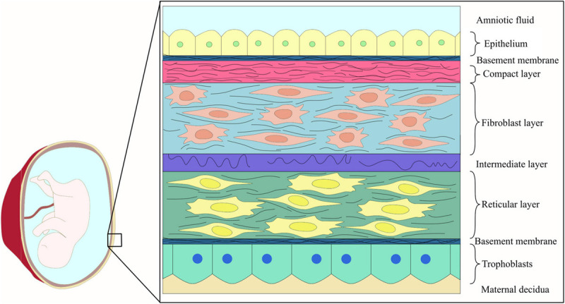
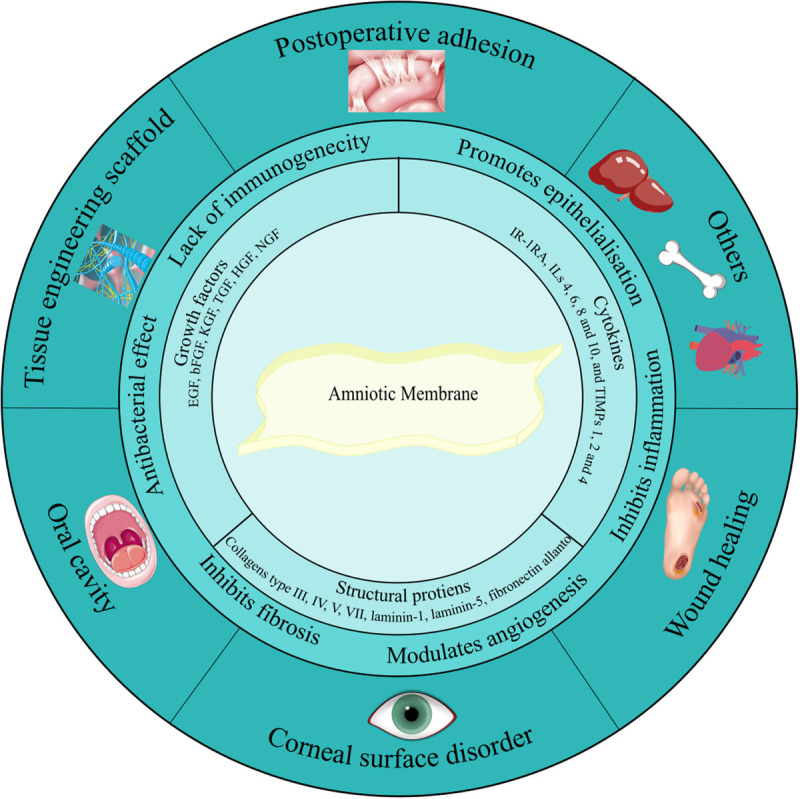
The amniotic membrane (AM) is the innermost layer of the fetal placenta, which surrounds and protects the fetus. Its unique structure, in addition to its physical and biological properties, makes it a useful substance in many applications related to regenerative medicine. The use of this fantastic substance with a century-old history has produced remarkable results in vivo, in vitro, and even in clinical studies. While the intact or preserved AM is widely used for these purposes, the addition of further modifications to AM can be considered as a relatively new subject in its applications. These modifications are applied to improve AM properties, ease of handling, and durability. Here, we will discuss the cases in which AM has undergone additional modifications besides the required processes for sterilization and preservation. In this article, we have categorized these modifications and discussed their applications and results.

Keywords: amniotic membrane; composites; hydrogel; regenerative medicine; tissue engineering.


Conflict of interest statement

The authors declare that the research was conducted in the absence of any commercial or financial relationships that could be construed as a potential conflict of interest.

Figures

FIGURE 1
FIGURE 1
Schematic representation of fetal membrane structure.
FIGURE 2
FIGURE 2
Amniotic membrane components, characteristics, and applications. Amniotic membrane is rich in growth factors, cytokines, and structural proteins. These biological factors have given AM its unique features such as antibacterial effect, anti-inflammatory activity, anti-scarring, and anti-fibrosis potential. Due to these suitable features, nowadays AM is widely utilized as a therapeutic option in the treatment of various diseases related to the urinary tract, oral cavity, skin, stomach, larynx, head and neck, ocular surface, pelvic, and abdominal surgery. Additionally, it has been used as a natural scaffold in TE.
FIGURE 3
FIGURE 3
(A) Schematic representation of direct electrospinning of secondary material on AM. (B) Conjugation of the surface-activated nanofiber mesh on the AM. (C) Gross healed wound areas after 30 days of wound healing from front and back views comparing AM and composite effect [reproduced with permission from Mandal et al., 2017. Copyright 2020 Elsevier]. (D) Maintenance of structural integrity, reduction of vascularization, and degradation after PCL-dAM composite transplantation in comparison with the AM-treated group for treating alkali-burn induced LSCD model [reproduced with permission from Zhou et al., 2019. Copyright 2019 Elsevier]. (E) Attachment and infiltration of Wharton’s jelly-derived MSCs seeded on the PLLA scaffolds after 7 days in two different magnification. (1, 2) Aligned PLLA scaffold containing ASA. (3, 4) Aligned PLLA scaffold containing ASA and coated with AM lysate [reproduced with permission from Aslani et al., 2019. Copyright 2019 Wiley]. (F) Representative series of expression of corneal epithelium-specific keratin 3 (K3) in epithelial cells cultured on (1) PVA-AM, (2) PVA-collagen, and (3) normal rabbit cornea [reproduced with permission from Uchino et al., 2007. Copyright 2007 Wiley].
FIGURE 4
FIGURE 4
(A) Schematic representation of AME preparation. (B) Results of linear mechanical abrasion test performed on control and AME-treated HCE cells after 72 h. After 72 h of mechanical abrasion, confluency was reached in AME-treated plates showing the healing effect of AME on mechanical cellular injury. Confluence is noted by the asterisk [reproduced with permission from Dudok et al., 2015. Copyright 2015 Wiley]. (C) Proof of reduction of ocular surface abnormalities induced by BAC with AE. The dry eye group was left untreated, and 200 μg/ml AE was administered for the AE200-treated group 3 times daily. On the 6th day, hyperemia was evaluated, and fluorescein staining and H&E staining were also performed. The white arrow in (2) shows ciliary hyperemia in dry eye condition, that in (8) shows the epithelium was weakened, and the other one shows the infiltration cell. Corneas treated with AE had smoother epithelium and less inflammation [Reproduced with permission from Xiao et al., 2013. Copyright 2013 Elsevier].
FIGURE 5
FIGURE 5
(A) Schematic representation of the preparation of AM hydrogel alone or in combination with a secondary substrate. (B) Histological images with H&E staining. Comparison of the efficiency of AM powder/hydrogel with commercially available products and other control groups. Skin treated with AM hydrogel and AM powder were very similar to the healthy skin [reproduced with permission from Tseng, 2016. Copyright 2019 Wiley]. (C) Placement of the AMED in the injured site of the fetal membrane by minimally invasive fetoscopic surgery [reproduced with permission from Lee et al., 2018. Copyright 2018 Wiley].

References

    1. Adamowicz J., Juszczak K., Bajek A., Tworkiewicz J., Nowacki M., Marszalek A., et al. (2012). Morphological and urodynamic evaluation of urinary bladder wall regeneration: muscles guarantee contraction but not proper function—a rat model research study. Transplant. Proc. 44 1429–1434. 10.1016/j.transproceed.2012.01.144 - DOI - PubMed
    1. Adamowicz J., Pasternak I., Kloskowski T., Gniadek M., Van Breda S. V., Buhl M., et al. (2020). Development of a conductive biocomposite combining graphene and amniotic membrane for replacement of the neuronal network of tissue-engineered urinary bladder. Sci. Rep. 10 1–15. - PMC - PubMed
    1. Adamowicz J., Pokrywczyńska M., Tworkiewicz J., Kowalczyk T., van Breda S. V., Tyloch D., et al. (2016). New amniotic membrane based biocomposite for future application in reconstructive urology. PLoS One 11:e0146012. 10.1371/journal.pone.0146012 - DOI - PMC - PubMed
    1. Akyürek M., Orhan E., Aydın M.Ş, Uysal Ö, Karşıdağ S. (2020). The effects of human amniotic membrane on silicone related capsule formation in rats. J. Plast. Surg. Hand Surg. 54 284–289. 10.1080/2000656x.2020.1766476 - DOI - PubMed
    1. Alamouti M. A., Shayan N., Momeni M., Koohkan A., Hajizadeh-Saffar E., Nouri M., et al. (2019). Investigation on the safety of amniotic membrane extracts in improving diabetic foot ulcers (phase 1 clinical trial study). Iran. J. Diab. Metabol. 18 0–0.