. 2021 Jan 14:8:575903.
doi: 10.3389/fcell.2020.575903. eCollection 2020.

MICAL-L2 Is Essential for c-Myc Deubiquitination and Stability in Non-small Cell Lung Cancer Cells

Affiliations

MICAL-L2 Is Essential for c-Myc Deubiquitination and Stability in Non-small Cell Lung Cancer Cells

Pengxiang Min et al. Front Cell Dev Biol. .

Abstract

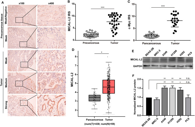
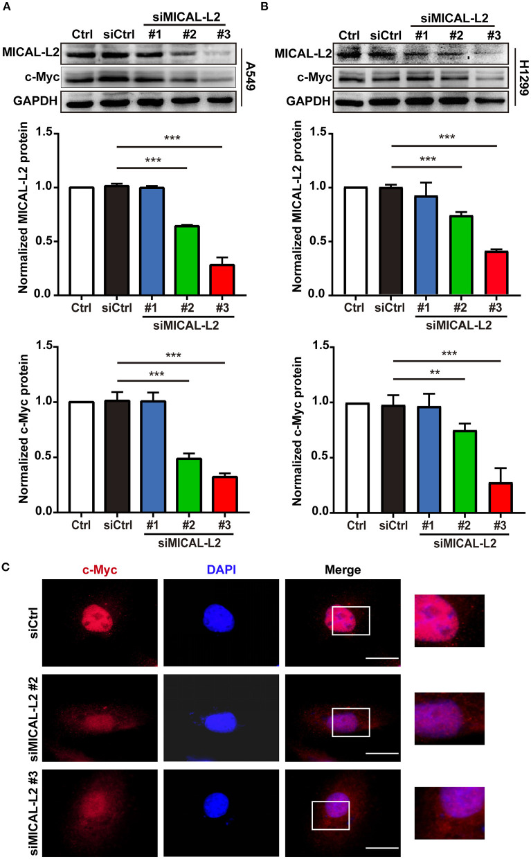
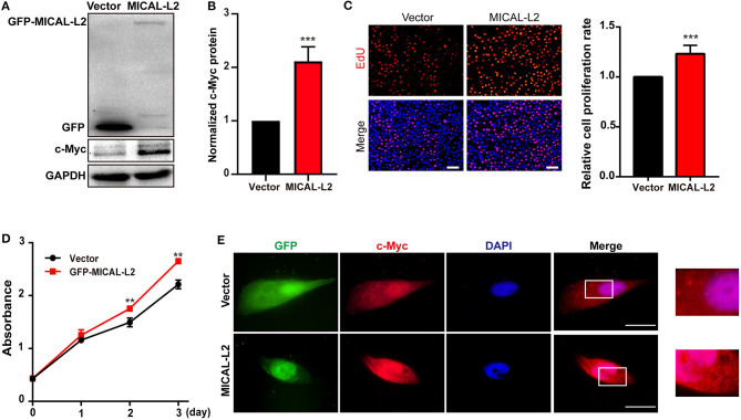
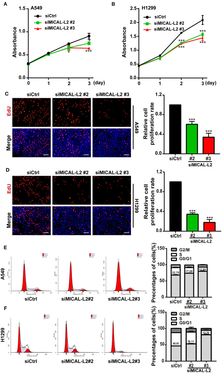
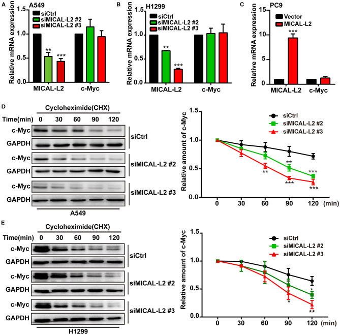
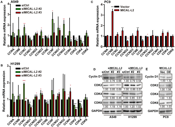
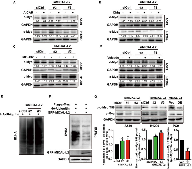
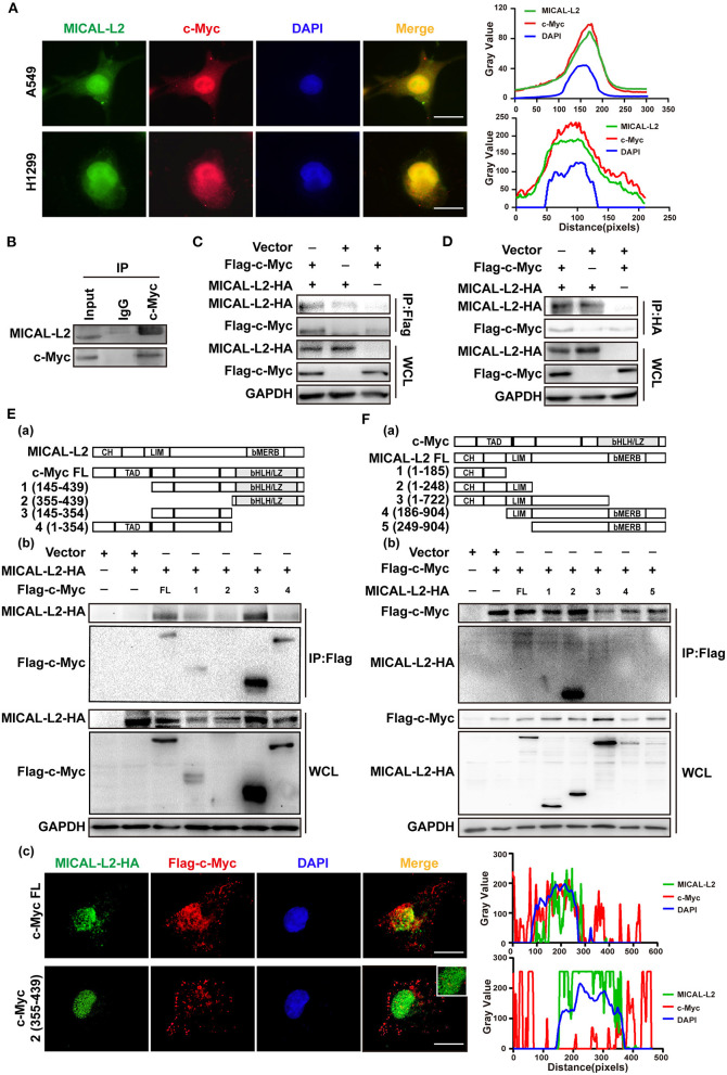
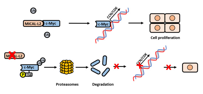
Objectives: MICAL-L2, a member of the molecules interacting with the CasL (MICAL) family, was reported to be highly expressed in several types of cancers, however, the roles of MICAL-L2 in NSCLC pathogenesis remain to be explored. This study is designed to clarify the mechanisms by which MICAL-L2 participates in NSCLC cell proliferation. Materials and Methods: The expression levels of MICAL-L2 in human lung cancer samples were assessed by immunohistochemical staining. Cells were transfected with siRNA or plasmids to regulate MICAL-L2 expression. Cell proliferation was measured by EdU staining and CCK-8 assays. MICAL-L2 and phosphorylated/total c-Myc expression were examined by Western blotting analysis. Interaction between MICAL-L2 and c-Myc was assessed by immunofluorescence staining, Western blotting and co-immunoprecipitation assays. Western blotting, polyubiquitylation detection and protein stability assays were used to assess whether MICAL-L2 exerts its oncogenic effect via c-Myc. Results: We found that MICAL-L2 was highly expressed in human NSCLC. While overexpressing MICAL-L2 increased NSCLC cell proliferation, MICAL-L2 depletion decreased the proliferation of NSCLC cells, an effect that was linked to cell cycle arrest. MICAL-L2 physically interacted with the c-Myc protein and functioned to maintain nuclear c-Myc levels and prolonged its half-life. Knockdown of MICAL-L2 expression led to decreased c-Myc protein stability through accelerating polyubiquitylation of c-Myc and gave rise to c-Myc degradation. We further found that MICAL-L2 deubiquitinated c-Myc and blocked its degradation, presumably by inhibiting c-Myc phosphorylation at threonine residue 58. Conclusions: These results indicate that MICAL-L2 is a key regulator of c-Myc deubiquitination and stability in the nucleus, and this activity may be involved in promoting NSCLC cell proliferation.

Keywords: MICAL-L2; NSCLC; c-Myc; deubiquitination; proliferation.


Conflict of interest statement

The authors declare that the research was conducted in the absence of any commercial or financial relationships that could be construed as a potential conflict of interest.

Figures

Figure 1
Figure 1
Analysis of MICAL-L2 and c-Myc expression in lung adenocarcinoma (LUAD) tissues. (A) Immunohistochemical staining for MICAL-L2 in LUAD tissues. (B,C) Analysis of MICAL-L2 and c-Myc staining in LUAD tissues. (D) Analysis of The Cancer Genome Atlas (TCGA) database showed that MICAL-L2 is highly expressed in lung cancer tissues when compared with normal tissues. (E,F) MICAL-L2 protein expression in different non-small cell lung carcinoma (NSCLC) cell lines. *P < 0.05, **P < 0.01, ***P < 0.001.
Figure 2
Figure 2
The effects of MICAL-L2 knockdown on c-Myc expression in non-small cell lung carcinoma (NSCLC) cells. (A) Total protein extracts from A549 cells treated with small interfering RNAs targeting MICAL-L2 (siMICAL-L2) for 48 h were assessed for MICAL-L2 and c-Myc expression. ***P < 0.001 relative to cells expressing control siRNA. (B) Blots showing the protein expression of MICAL-L2 and c-Myc in lysates from H1299 cells transfected with siMICAL-L2. **P < 0.01, ***P < 0.001 relative to cells expressing control siRNA. Data in (A) and (B) are presented as means ± SD of 3 determinations. (C) Representative immunofluorescence images of c-Myc staining in A549 cells transfected with siMICAL-L2. Scale bar, 5 μm.
Figure 3
Figure 3
MICAL-L2 overexpression enhanced the proliferative ability of human non-small cell lung carcinoma (NSCLC) cells. (A,B) Total protein extracts from PC9 cells transfected with MICAL-L2 expression plasmids for 48 h were assessed for c-Myc expression. Data are presented as means ± SD of 3 determinations. ***P < 0.001 relative to control cells. (C) EdU staining in MICAL-L2-overexpressing PC9 cells. Data are presented as means ± SD of 5 independent determinations. (D) The viability of PC9 cells transfected with empty vector or MICAL-L2 expression plasmids was measured by CCK-8 assay. Data are presented as means ± SD of 10 determinations. (E) Representative immunofluorescence images of c-Myc staining in PC9 cells transfected with MICAL-L2 expression plasmids. Scale bar, 5 μm.**P < 0.01, ***P < 0.001 relative to control cells.
Figure 4
Figure 4
MICAL-L2 knockdown decreased the growth of non-small cell lung carcinoma (NSCLC) cells and induced S-phase cell cycle arrest. (A,B) CCK-8 assays for the viability of MICAL-L2-depleted A549 cells (left) and H1299 cells (right). (C,D) EdU staining showing the effect of MICAL-L2 depletion on the proliferative ability of A549 cells (C) and H1299 cells (D). (E,F) A549 cells (E) and H1299 cells (F) underwent cell cycle distribution analysis by flow cytometry. Cell cycle distribution data are shown in histograms. *P < 0.05, ***P < 0.001 relative to control cells.
Figure 5
Figure 5
MICAL-L2 reduced c-Myc degradation. (A,B) The mRNA levels of MICAL-L2 and c-Myc were determined by RT-qPCR in A549 cells (left) and H1299 cells (right) transfected with small interfering (si) RNAs targeting MICAL-L2 (siMICAL-L2) and (C) PC9 cells transfected with MICAL-L2 expression plasmids. (D,E) The protein levels of c-Myc were examined in A549 and H1299 cells transfected with control siRNA or siMICAL-L2 and treated with cycloheximide (CHX, 10 μg/mL) for the indicated times. *P < 0.05, **P < 0.01, ***P < 0.001 relative to cells expressing control siRNA.
Figure 6
Figure 6
The effect of MICAL-L2 on cell cycle-related proteins. (A,B) RT-qPCR analysis of CDK2, CDK4, CDK6, and CCNA-H mRNA levels in A549 and H1299 cells transfected with control small interfering (si) RNA or siRNA targeting MICAL-2 (siMICAL-2). *P < 0.05, **P < 0.01, ***P < 0.001 relative to cells expressing control siRNA. (C) RT-qPCR analysis of CDK2, CDK4, CDK6, and CCNA-H mRNA levels in A549 cells transfected with empty vectors or MICAL-L2 expression plasmids. (D,E) Western blotting analysis of cyclin-D1, CDK2, CDK4, and CDK6 protein levels in MICAL-L2-depleted (D) or MICAL-L2-overexpressing (E) A549 cells. *P < 0.05, **P < 0.01, ***P < 0.001 relative to control cells.
Figure 7
Figure 7
MICAL-L2 inhibited c-Myc ubiquitin-mediated degradation. (A–D) A549 and H1299 cells transfected with small interfering (si) RNA targeting MICAL-L2 (siMICAL-L2) were treated with AICAR (0.2 mM), chloroquine (10 μM), MG-132 (20 μM), and Velcade (10 μM) for 24 h. Total proteins were then extracted from the lysates and subjected to Western blotting to detect the expression of c-Myc. GAPDH served as the loading control. (E) H1299 cells were transfected with HA-ubiquitin and siMICAL-L2 and c-Myc polyubiquitination was detected by Western blotting using the indicated antibodies. (F) H1299 cells were co-transfected with HA-ubiquitin and GFP-MICAL-L2, Flag-c-Myc, or empty vector, following which c-Myc polyubiquitination was assayed. (G) A549 and H1299 cells were transfected with control siRNA or siMICAL-2. Total proteins were then extracted and analyzed for the expression of phosphorylated-c-Myc (T58) by Western blotting. PC9 cells were transfected with empty vector or MICAL-L2 expression plasmids. Total proteins were then extracted and analyzed for the expression of phosphorylated-c-Myc (T58) by Western blotting. Western blotting bands corresponding to phosphorylated-c-Myc/c-Myc were quantified and normalized against GAPDH. *P < 0.05, **P < 0.01 relative to control cells.
Figure 8
Figure 8
MICAL-L2 interacted with c-Myc. (A) Representative immunofluorescence images of MICAL-L2 (green), c-Myc (red), and nuclei (blue) staining in A549 and H1299 cells. (B) The binding of endogenous MICAL-L2 to c-Myc was detected in H1299 cells by co-immunoprecipitation assays. (C,D) Co-immunoprecipitation was performed with extracts from Cos-7 cells co-transfected with Flag-tagged c-Myc and HA-tagged MICAL-L2. (E) Schematic representation of the c-Myc domains (a). Cos-7 cells were co-transfected with HA-MICAL-L2 and a c-Myc mutant following which cell extracts were analyzed by Western blotting (b). Cos-7 cells were co-transfected with HA-MICAL-L2 and c-Myc mutant #2 following which cell extracts were analyzed using Immunofluorescence (c). (F) Schematic representation of the MICAL-L2 domains (a). Cos-7 cells were co-transfected with Flag-c-Myc and a MICAL-L2 mutant following which cell extracts were analyzed by Western blotting (b).
Figure 9
Figure 9
Schematic model for how MICAL-L2 regulates c-Myc expression and c-Myc-mediated cell proliferation. In brief, MICAL-L2 was identified as a novel key molecule implicated in non-small cell lung carcinoma (NSCLC) cell proliferation. MICAL-L2 binds to c-Myc, thereby suppressing c-Myc ubiquitination and degradation, which increases the expression of c-Myc target genes (including cell cycle-related genes) to promote NSCLC cell proliferation.

Similar articles

Cited by

References

    1. Agrawal P., Yu K., Salomon A. R., Sedivy J. M. (2010). Proteomic profiling of Myc-associated proteins. Cell Cycle 9, 4908–4921. 10.4161/cc.9.24.14199 - DOI - PMC - PubMed
    1. Cai Y., Lu J., Tang F. (2018). Overexpression of MICAL2, a novel tumor-promoting factor, accelerates tumor progression through regulating cell proliferation and EMT. J. Cancer 9, 521–527. 10.7150/jca.22355 - DOI - PMC - PubMed
    1. Dang C. V. (1999). c-Myc target genes involved in cell growth, apoptosis, and metabolism. Mol. Cell. Biol. 19, 1–11. 10.1128/MCB.19.1.1 - DOI - PMC - PubMed
    1. Dang C. V. (2012). MYC on the path to cancer. Cell 149, 22–35. 10.1016/j.cell.2012.03.003 - DOI - PMC - PubMed
    1. Deng W., Wang Y., Gu L., Duan B., Cui J., Zhang Y., et al. . (2016). MICAL1 controls cell invasive phenotype via regulating oxidative stress in breast cancer cells. BMC Cancer 16:489. 10.1186/s12885-016-2553-1 - DOI - PMC - PubMed

LinkOut - more resources