

Etiological profile and main imaging findings in patients with granulomatous diseases who underwent lung biopsy
- PMID: 33521170
- PMCID: PMC7820493
- DOI: 10.1016/j.ejro.2021.100325
Etiological profile and main imaging findings in patients with granulomatous diseases who underwent lung biopsy
Abstract
Background: Granulomatous Lung Diseases (GLD) encompasses a wide range of infectious and non-infectious conditions characterized by chronic inflammatory response. However, different GLD may share similar imaging findings. In this context, the purpose of this study was to outline the etiological profile and their imaging features in patients with GLD who underwent lung biopsy.
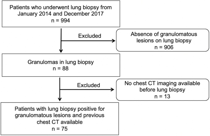
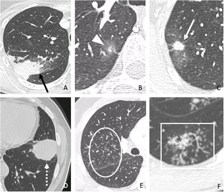
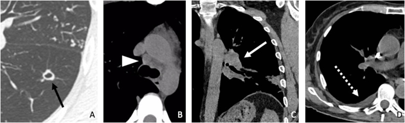
Methods: Patients with granulomatous lesions in lung biopsies and previous chest CT performed from 2014 to 2017 at our institution had imaging data reviewed by three blinded radiologists. The imaging features were analyzed according to the Fleischner Society glossary. Categorical data were represented by absolute (n) and relative (%) frequency. The contingency matrices were analyzed by Pearson's Chi-square test. Interreader agreement was assessed by calculating the intraclass correlation coefficient, using kappa (κ) statistic.
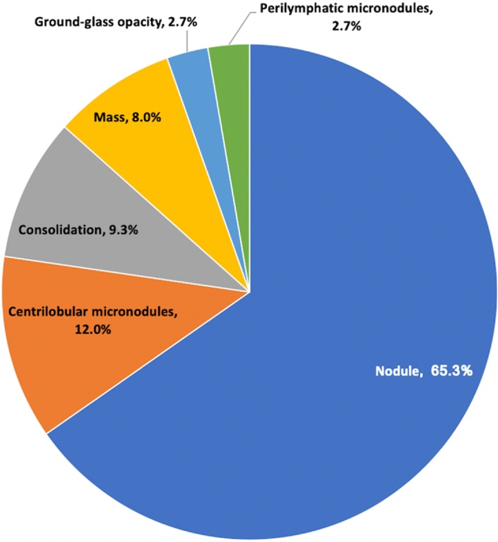
Results: Thirty-eight of 75 (50.7%) patients were women with a mean age of 59 ± 39 years. Infection was the most common cause of GLD (47/75, 62.7%) and Histoplasma capsulatum (27/75, 36%) was the most prevalent etiology. Nodular pattern was the most common imaging feature in histoplasmosis cases (25/27, 92.6%), whereas it occurred in half of cases (24/48) of GLD of other causes (p < 0.05). Among patients with tuberculosis, the second etiology of GLD in our study population, the most common imaging pattern was centrilobular micronodules (3/7, 42.9%), significantly more frequent than in other causes of GLD (6/68, 8.8%). Interreader agreement in detecting imaging features was almost perfect (κ = 0.88-1.00), except the nodular pattern, which had substantial agreement (κ = 0.73).
Conclusions: In our study population, the main etiologies found in patients with granulomatous disease who underwent lung biopsy were fungal or mycobacterial disease, specially histoplasmosis and tuberculosis, and nodular pattern with focal distribution was the most common imaging finding which was detected with substantial interreader agreement.
Keywords: CT, Computed Tomography; FOV, Field of View; GLD, Granulomatous lung diseases; GLUS, Granulomatous Lesions of Unknown Significance; Granuloma; JR, Junior Radiologists; TB, Tuberculosis; histoplasmosis; lung biopsy; pulmonary tuberculosis; solitary pulmonary nodule; κ, Kappa.
© 2021 The Authors.
Figures
References
-
- Zhu Q., Xu X., Li M., Wang X. Analysis of chest computed tomography manifestations of non-Mycobacterium tuberculosis induced granulomatous lung diseases. Radiology of Infectious Diseases. 2017:157–163.
-
- Yang X., He J., Wang J., Li W., Liu C., Gao D. CT-based radiomics signature for differentiating solitary granulomatous nodules from solid lung adenocarcinoma. Lung Cancer. 2018;125:109–114. - PubMed
-
- Nachiappan A.C., Rahbar K., Shi X., Guy E.S., Mortani Barbosa E.J., Shroff G.S. Pulmonary Tuberculosis: Role of Radiology in Diagnosis and Management. Radiographics. 2017;37(1):52–72. - PubMed
-
- WHO . World Health Organization; Geneva: 2019. Global tuberculosis report 2019.
Grants and funding
LinkOut - more resources
Full Text Sources
Other Literature Sources
