Review
. 2021;5(3):197-216.
doi: 10.1038/s41570-020-00244-3. Epub 2021 Jan 25.

Natural and synthetic carbohydrate-based vaccine adjuvants and their mechanisms of action

Affiliations
Review

Natural and synthetic carbohydrate-based vaccine adjuvants and their mechanisms of action

Carlo Pifferi et al. Nat Rev Chem. 2021.

Abstract

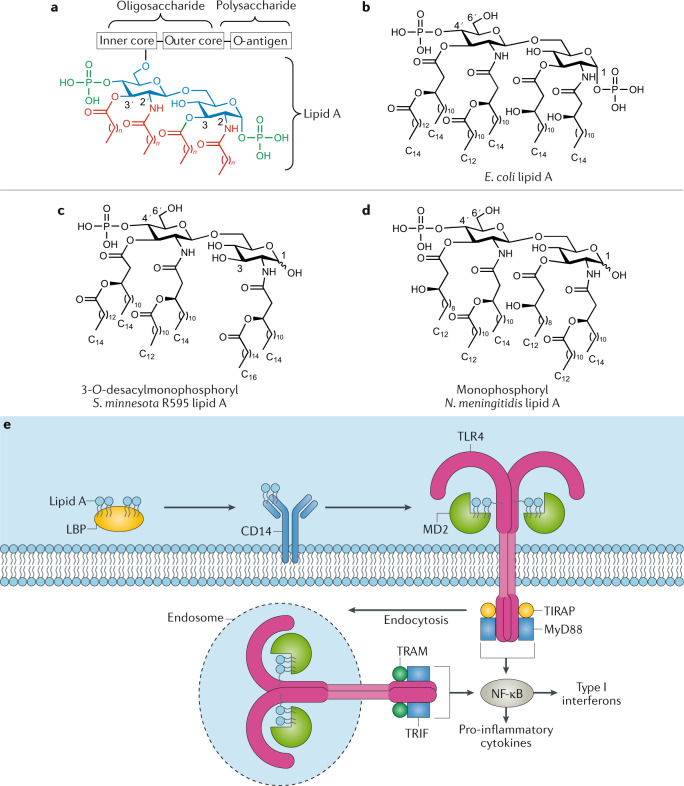
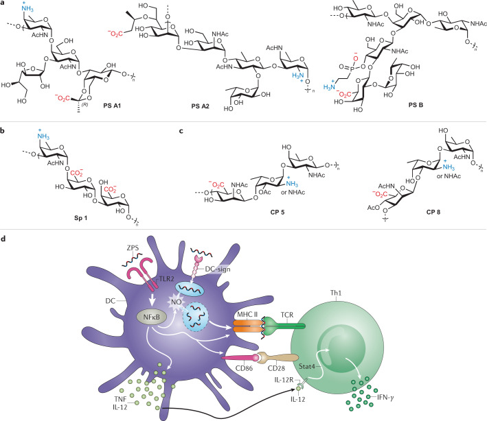
Modern subunit vaccines based on homogeneous antigens offer more precise targeting and improved safety compared with traditional whole-pathogen vaccines. However, they are also less immunogenic and require an adjuvant to increase the immunogenicity of the antigen and potentiate the immune response. Unfortunately, few adjuvants have sufficient potency and low enough toxicity for clinical use, highlighting the urgent need for new, potent and safe adjuvants. Notably, a number of natural and synthetic carbohydrate structures have been used as adjuvants in clinical trials, and two have recently been approved in human vaccines. However, naturally derived carbohydrate adjuvants are heterogeneous, difficult to obtain and, in some cases, unstable. In addition, their molecular mechanisms of action are generally not fully understood, partly owing to the lack of tools to elucidate their immune-potentiating effects, thus hampering the rational development of optimized adjuvants. To address these challenges, modification of the natural product structure using synthetic chemistry emerges as an attractive approach to develop well-defined, improved carbohydrate-containing adjuvants and chemical probes for mechanistic investigation. This Review describes selected examples of natural and synthetic carbohydrate-based adjuvants and their application in synthetic self-adjuvanting vaccines, while also discussing current understanding of their molecular mechanisms of action.

Keywords: Adjuvants; Carbohydrate chemistry; Carbohydrates; Chemical modification; Mechanism of action.


Conflict of interest statement

Competing interestsA.F.-T. is co-inventor on patents and patent applications that include synthetic QS-21 variants mentioned in this work. C.P. and R.F. declare no competing interests.

Figures

Fig. 1
Fig. 1. Structures of natural and synthetic QS-based saponin adjuvants and proposed mechanism of action for QS-21-related saponin adjuvants.
a | Structures of saponin natural product adjuvants QS-21, QS-18 and QS-17 derived from the Quillaja saponaria tree and summary of structure–adjuvant activity relationships of QS-21 (ref.). b | Structures of saponin natural product adjuvant QS-7Xyl (ref.) and summary of QS-7 structure–adjuvant activity relationships,. c | Schematic representation of the proposed mechanism of action for QS-21-related saponin adjuvants. Upon endocytosis, exogenous protein antigens and QS-21 are delivered to dendritic cells (DCs). Following QS-21-mediated disruption of the endosomal membrane, cleaved protein antigens can be further processed into smaller peptide fragments in the cytosol by the proteasome machinery. Degraded peptides are translocated into the endoplasmic reticulum (ER) by transporter molecules, where chaperones facilitate their binding to newly synthesized MHC class I (MHC-I) molecules for vesicular migration through the Golgi to the cell surface. Finally, peptide epitopes exposed on the DC surface in association with MHC-I molecules are presented to naive CD8+ T cells (cross-presentation) through the T cell receptor (TCR). TH, T helper. Part c adapted from ref., CC BY 4.0 (https://creativecommons.org/licenses/by/4.0/), and with permission from ref., Elsevier.
Fig. 2
Fig. 2. Structures of natural and synthetic α-GalCer-based adjuvants and mechanism of action of α-GalCer.
a | Structure of natural α-galactosylceramide (α-GalCer) and key structural modifications and their impact on activity and cytokine profile production. b,c | Structures of synthetic α-GalCer variants inducing T helper 2 (TH2)-biased (OCH, panel b) and TH1-biased (7DW8-5, panel c) responses. d | Schematic representation of invariant natural killer T (iNKT) cell activation and α-GalCer mechanism of action. α-GalCer presentation on antigen-presenting cell (APC) surface in association with CD1d enables activation of iNKT cells by interaction with their invariant T cell receptor (iTCR). iNKT cells rapidly secrete both pro-inflammatory (TH1) and anti-inflammatory (TH2) cytokines, such as interferon-γ (IFNγ) and interleukin-4 (IL-4), respectively. Depending on several factors, including the nature of the glycolipid antigen, its loading mode into CD1d protein, cytokine milieu, cell types that present the glycolipid antigen, co-stimulatory interactions and frequency of treatment, activated iNKT cells can exhibit a diverse range of responses on other cell types, such as B and T lymphocytes, macrophages, dendritic cells and NK cells. Part d adapted with permission from ref., ACS, from ref., CC BY 4.0 (https://creativecommons.org/licenses/by/4.0/), and with permission from ref., Future Medicine.
Fig. 3
Fig. 3. Structures of lipid A and monophosphoryl lipid A and its mechanism of action.
a | General structure of lipopolysaccharide (LPS) with its three main domains, lipid A, core oligosaccharide and O-antigen polysaccharide. b | Structure of natural lipid A from Escherichia coli. c,d | Structures of chemically modified monophosphoryl lipid A from Salmonella minnesota R595 (clinically approved, panel c) and from Neisseria meningitidis (panel d). e | Schematic representation of LPS/Toll-like receptor 4 (TLR4) signalling pathway and lipid A mechanism of action. LPS-binding protein (LBP)-associated LPS (or lipid A) is first transferred to CD14 and then delivered to myeloid differentiation factor 2 (MD2), enabling formation of the LPS–MD2–TLR4 ternary complex, which drives dimerization of TLR4 receptors. LPS/TLR4 signalling involves intracellular recruitment of Toll/interlukin-1 (IL-1) receptor domain-containing adapter protein (TIRAP), TRIF-related adapter molecule (TRAM), myeloid differentiation factor 88 (MyD88) and Toll/IL-1 receptor domain-containing adapter inducing interferon-β (TRIF) co-receptors. While the MyD88-dependent pathway promotes pro-inflammatory cytokine expression, the MyD88-independent pathway mediates the induction of type I interferons. Part e adapted with permission from ref., Wiley, and with permission from ref., Elsevier.
Fig. 4
Fig. 4. Structures of natural zwitterionic polysaccharides and their proposed mechanism of action.
a | Structures of natural zwitterionic polysaccharides (ZPSs) PS A1, PS A2 and PS B from Bacteroides fragilis. b,c | Structures of natural ZPSs from Streptococcus pneumoniae (type 1, Sp 1, panel b) and Staphylococcus aureus (type 5, CP5, and type 8, CP8, panel c). d | Schematic representation of the proposed model for ZPS mechanism of action and crosstalk between innate and adaptive immune compartments. Within the innate immunity context (left), Toll-like receptor 2 (TLR2)-mediated recognition of zwitterionic polysaccharide induces the activation of the myeloid differentiation factor 88 (MyD88)-dependent pathway in antigen-presenting cells (e.g. dendritic cells), leading to NF-κB-dependent pro-inflammatory cytokine expression (tumour necrosis factor (TNF), interleukin-12 (IL-12)), NO production, and MHC class II (MHC-II) and co-stimulatory molecule expression and upregulation. Adaptive immunity events (right) involving interaction between T cell receptor (TCR) and processed ZPSs presented on MHC-II, along with co-stimulation via CD86/CD28 and IL-12/IL-12R interactions, ultimately trigger ZPS-activated CD4+ T cells to produce the T helper 1 (TH1) cytokine interferon-γ (IFNγ). DC, dendritic cell. Part d adapted with permission from ref., Rockefeller University Press.

References

    1. Jones LH. Recent advances in the molecular design of synthetic vaccines. Nat. Chem. 2015;7:952–960. doi: 10.1038/nchem.2396. - DOI - PubMed
    1. Reed SG, Orr MT, Fox CB. Key roles of adjuvants in modern vaccines. Nat. Med. 2013;19:1597–1608. doi: 10.1038/nm.3409. - DOI - PubMed
    1. Flach TL, et al. Alum interaction with dendritic cell membrane lipids is essential for its adjuvanticity. Nat. Med. 2011;17:479–487. doi: 10.1038/nm.2306. - DOI - PubMed
    1. HogenEsch H, O’Hagan DT, Fox CB. Optimizing the utilization of aluminum adjuvants in vaccines: you might just get what you want. NPJ Vaccines. 2018;3:51. doi: 10.1038/s41541-018-0089-x. - DOI - PMC - PubMed
    1. Del Giudice G, Rappuoli R, Didierlaurent AM. Correlates of adjuvanticity: a review on adjuvants in licensed vaccines. Semin. Immunol. 2018;39:14–21. doi: 10.1016/j.smim.2018.05.001. - DOI - PubMed