. 2021 Feb 19;24(2):102054.
doi: 10.1016/j.isci.2021.102054. Epub 2021 Jan 9.

Immunogenicity and protective efficacy of BBV152, whole virion inactivated SARS- CoV-2 vaccine candidates in the Syrian hamster model

Affiliations

Immunogenicity and protective efficacy of BBV152, whole virion inactivated SARS- CoV-2 vaccine candidates in the Syrian hamster model

Sreelekshmy Mohandas et al. iScience. .

Abstract

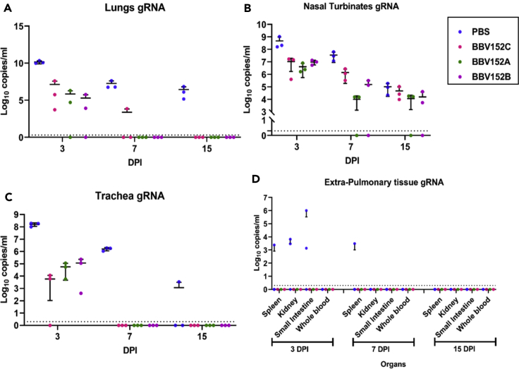
The availability of a safe and effective vaccine would be the eventual measure to deal with severe acute respiratory syndrome coronavirus-2 (SARS-CoV-2) threat. Here, we have assessed the immunogenicity and protective efficacy of inactivated SARS-CoV-2 vaccine candidates BBV152A, BBV152B, and BBV152C in Syrian hamsters. Three dose vaccination regimes with vaccine candidates induced significant titers of SARS-CoV-2-specific IgG and neutralizing antibodies. BBV152A and BBV152B vaccine candidates remarkably generated a quick and robust immune response. Post-SARS-CoV-2 infection, vaccinated hamsters did not show any histopathological changes in the lungs. The protection of the hamster was evident by the rapid clearance of the virus from lower respiratory tract, reduced virus load in upper respiratory tract, absence of lung pathology, and robust humoral immune response. These findings confirm the immunogenic potential of the vaccine candidates and further protection of hamsters challenged with SARS-CoV-2. Of the three candidates, BBV152A showed the better response.

Keywords: Biological Sciences; Immunity; Immunology; Viral Microbiology; Virology.


Conflict of interest statement

The authors declare no competing interests.

Figures

None
Graphical abstract
Figure 1
Figure 1
Schematic presentation of experiments in hamsters Three doses of placebo were administered to the first group of the animals which were controls in the study. Three different inactivated SARS-CoV-2 vaccine formulations (2 doses + boost) were administered to the three groups of animals. All the animals were challenged at 15 days after the third dose. Samples were collected at different time points of immunization period and post-infection as indicated in the figure. Necropsy was performed for three hamsters from each group at 3, 7, and 15 DPI.
Figure 2
Figure 2
Percent body weight gain/loss in hamsters (A) Percentage of body weight gain in hamsters during the immunization period. (B) Percent difference in body weight in hamsters after SARS-CoV-2 challenge. Mean along with standard deviation (SD) is depicted in the scatterplot. The statistical significance was assessed using the Kruskal-Wallis test followed by the two-tailed Mann-Whitney test between the two groups; p values less than 0.05 were considered to be statistically significant.
Figure 3
Figure 3
Humoral response in vaccinated animals (A) IgG antibody response for all groups of animals observed on 12, 21, and 48 days. (B) IgG antibody response at post-infection (3, 7, and 15 DPI) for all groups of animals. (C) Comparison of IgG antibody titers between groups on 12, 21, and 48 days. (D) Comparison of IgG antibody titers between groups after virus challenge at 3, 7, and 15 DPI. (E). Comparison of IgG2 antibody titers between groups during immunization period at 21 and 48 days and post virus challenge at 3,7, and 15 DPI. (F and G) (F). Comparison of NAb titers response during a three-dose vaccine regime for all groups of animals observed on 12, 21, and 48 days (G). Comparison of NAb titers response in SARS-CoV-2 infected animals on 3, 7, and 15 DPI. Mean along with standard deviation (SD) is depicted in the scatterplot. The statistical significance was assessed using the Kruskal-Wallis test followed by the two-tailed Mann-Whitney test between the two groups; p values less than 0.05 were considered to be statistically significant. The dotted lines indicate the limit of detection of the assay.
Figure 4
Figure 4
Log10 plot for the genomic viral RNA detection in throat swab and nasal wash after virus challenge Genomic viral RNA load in (A) Throat Swab collected at 3, 5, 7, 10, and 15 DPI for the all groups (B) Nasal wash genomic viral RNA at 3, 7, and 15 DPI for the all groups. Mean along with standard deviation (SD) is depicted in the scatterplot. The statistical significance was assessed using the Kruskal-Wallis test followed by the two-tailed Mann-Whitney test between the two groups; p values less than 0.05 were considered to be statistically significant. The dotted lines indicate the limit of detection of the assay.
Figure 5
Figure 5
Log10 plot of the genomic viral RNA detection in the respiratory tract and extra pulmonary specimens Genomic viral RNA load in (A) lung, (B) nasal turbinates, (C) trachea, (D) extra pulmonary organs at 3, 7, and 15 DPI. Mean along with standard deviation (SD) is depicted in the scatterplot. The statistical significance was assessed using the Kruskal-Wallis test followed by the two-tailed Mann-Whitney test between the two groups; p values less than 0.05 were considered to be statistically significant. The dotted lines indicate the limit of detection of the assay.
Figure 6
Figure 6
Gross and histopathological observations of lungs in hamsters after virus inoculation (A–D) (A) Lungs of hamster from group I on 7 DPI showing diffuse areas of consolidation and congestion in the left and right lower lobe with few congestive foci in right upper lobe, scale bar = 0.73cm. Lungs from (B) group II, scale bar = 0.65cm (C) group III, scale bar = 0.73cm and (D) group IV showing normal gross appearance on 7 DPI scale bar = 0.52cm. (E) Lung tissue from group I on 3 DPI showing acute inflammatory response with diffuse alveolar damage, haemorrhages, inflammatory cell infiltration (black arrow), hyaline membrane formation (white arrow), and accumulation of eosinophilic edematous exudate (star), scale bar = 20μm. (F) Lung tissue of group I on 7 DPI showing acute interstitial pneumonia with marked alveolar damage, thickening of alveolar and accumulation of mononuclear cells, and macrophages (white arrow), and lysed erythrocytes in the alveolar luminal space (star), scale bar = 20μm. (G–J) (G) Lung tissue from group I on 15 DPI depicting interstitial pneumonia with marked thickening of alveolar septa with type-II pneumocyte hyperplasia and fibro-elastic proliferation with collagen deposition at alveolar epithelial lining (white arrow), scale bar = 20μm. Lung section from group II showing no evidence of disease (H) on 3 DPI few congestive foci, scale bar = 20μm (I) on 7 DPI, scale bar = 20μm (J) on 15 DPI, scale bar = 20μm.
Figure 7
Figure 7
Immunohistochemistry findings in lungs of hamsters after virus challenge Left panel depicts group treated with placebo and the right panel shows vaccinated group II animals. Lung section from group I showing viral antigen (A) on 3 DPI in alveolar type-II pneumocytes (black arrow) and in alveolar macrophages (white arrow), scale bar = 20μm (B) on 7 DPI in alveolar type-II pneumocytes (black arrow), scale bar = 20μm (C) on 15 DPI in type-II alveolar pneumocytes (black arrow), scale bar = 20μm. Lung section from group II showing viral antigen (D) on 3 DPI in alveolar macrophages (black arrow), scale bar = 20μm (E) on 7 DPI in alveolar epithelium and alveolar macrophages, scale bar = 20μm (F) on 15 DPI in the alveolar epithelium and alveolar macrophages, scale bar = 20μm.

References

    1. Addetia A., Crawford K.H., Dingens A., Zhu H., Roychoudhury P., Huang M.L., Jerome K.R., Bloom J.D., Greninger A.L. Neutralizing antibodies correlate with protection from SARS-CoV-2 in humans during a fishery vessel outbreak with a high attack rate. J. Clin. Microbiol. 2020;58:e02107–e02120. doi: 10.1128/JCM.02107-20. - DOI - PMC - PubMed
    1. Chan J.F., Zhang A.J., Yuan S., Poon V.K., Chan C.C., Lee A.C., Chan W.M., Fan Z., Tsoi H.W., Wen L., Liang R. Simulation of the clinical and pathological manifestations of Coronavirus Disease 2019 (COVID-19) in golden Syrian hamster model: implications for disease pathogenesis and transmissibility. Clin. Infect. Dis. Mar. 2020;26:ciaa325. - PMC - PubMed
    1. Clements J.D., Freytag L.C. Parenteral vaccination can be an effective means of inducing protective mucosal responses. Clin. Vaccin. Immunol. 2016;23:438–441. - PMC - PubMed
    1. Docea A.O., Tsatsakis A., Albulescu D., Cristea O., Zlatian O., Vinceti M., Moschos S.A., Tsoukalas D., Goumenou M., Drakoulis N., Dumanov J.M. A new threat from an old enemy: Re-emergence of coronavirus. Int. J. Mol. Med. 2020;45:1631–1643. - PMC - PubMed
    1. Ganneru B., Jogdand H., Dharam V.K., Molugu N.R., Prasad S.D., Vellimudu S., Ella K.M., Ravikrishnan R., Awasthi A., Jose J., Rao P. bioRxiv; 2020. Evaluation of Safety and Immunogenicity of an Adjuvanted, TH-1 Skewed, Whole Virion Inactivated SARS-CoV-2 Vaccine-Bbv152. - DOI