Review
. 2020 Dec:8:100043.
doi: 10.1016/j.medntd.2020.100043. Epub 2020 Aug 29.

2020 update on human coronaviruses: One health, one world

Affiliations
Review

2020 update on human coronaviruses: One health, one world

Xinbin Zhao et al. Med Nov Technol Devices. 2020 Dec.

Abstract

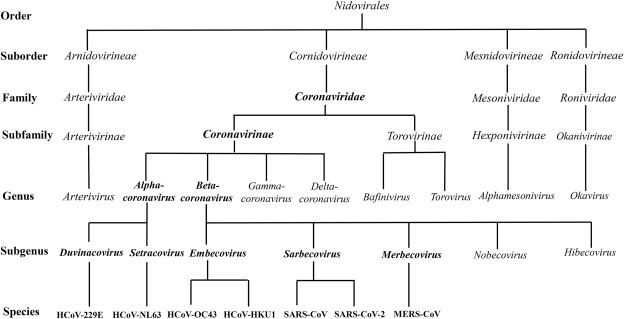
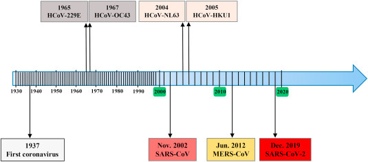
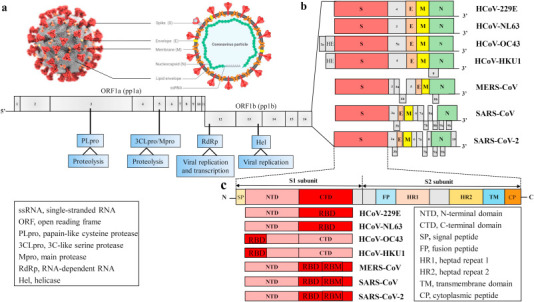
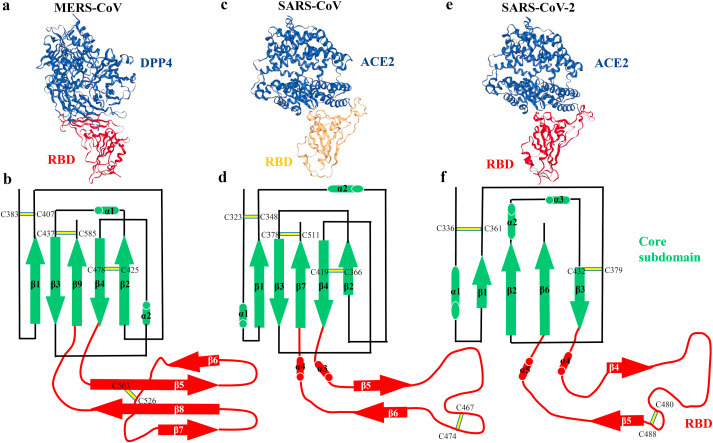
Since human coronavirus (HCoVs) was first described in the 1960s, seven strains of respiratory human coronaviruses have emerged and caused human infections. After the emergence of severe acute respiratory syndrome coronavirus (SARS-CoV) and Middle East respiratory syndrome coronavirus (MERS-CoV), a pneumonia outbreak of coronavirus disease 2019 (COVID-19) caused by a novel coronavirus (SARS-CoV-2) has represented a pandemic threat to global public health in the 21st century. Without effectively prophylactic and therapeutic strategies including vaccines and antiviral drugs, these three coronaviruses have caused severe respiratory syndrome and high case-fatality rates around the world. In this review, we detail the emergence event, origin and reservoirs of all HCoVs, compare the differences with regard to structure and receptor usage, and summarize therapeutic strategies for COVID-19 that cause severe pneumonia and global pandemic.

Keywords: Gene structure; Human coronavirus; Receptor usage; Reservoirs; Therapeutic strategies.


Figures

Fig. 1
Fig. 1
The taxonomy of the order Nidovirales. HCoV, human coronavirus; MERS, Middle East respiratory syndrome; SARS, severe acute respiratory syndrome.
Fig. 2
Fig. 2
Timeline for the emergency of human coronaviruses. HCoV, human coronavirus; MERS, Middle East respiratory syndrome; SARS, severe acute respiratory syndrome.
Fig. 3
Fig. 3
Genomes and structures of human coronaviruses. (a) The ultrastructural morphology of coronavirus on the left illustration (the Centers for Disease Control and Prevention, CDC) and right virus particle. (b) The genomes of HCoVs contain a single-stranded, positive-sense RNA (ssRNA) genome of 27–32 ​kb in size. The 5′-terminal ORF1a/b within two-thirds of the genome encodes two large polyproteins 1a (pp1a) and pp1b. These polyproteins are cleaved by PLpro and 3CLpro, also known as Mpro, to produce non-structural proteins (nsps), including RdRp and Hel, which are important enzymes involved in the transcription and replication. The 3′ one-third of genome encodes four structural proteins: Spike (S), Membrane (M), Envelope (E) and Nucleocapsid (N), which are essential for virus-cell receptor binding and virion assembly, and other non-structural proteins along with a set of accessory proteins unique to each virus species. Some coronaviruses express an additional structural protein, hemagglutinin-esterase (HE). (c) The S protein of HCoVs consists of S1 subunit and S2 subunit. The S1 region contains an NTD and a CTD (also referred to as the RBD), whereas the S2 region includes a TM region, FP, HR1 and HR2. The HE glycoprotein consists of two functional domains: a corresponding sialate O-acetylesterase domain and an O-acetylated sialic acid binding domain. HCoV-OC43 and HKU1 bind 9-O-Ac-Sia via NTD of S1 subunit and mediate sialate-O-acetylesterase activity by RDE domain of HE protein. Other coronaviruses mediate attachment to the cellular receptors by RBD (CTD) in S1 region. Herein, we compare and contrast genome structures of seven identified HCoVs strains HCoV-229 ​E, HCoV-NL63, HCoV-OC43, HCoV-HKU1, MERS-CoV, SARS-CoV and SARS-CoV-2.
Fig. 4
Fig. 4
The structural comparison of RBD bound with receptor and RBD subdomains in the three coronaviruses. (a) Overall structure of RBD bound with DPP4 (PDB ID: 4KR0), and (b) schematic illustration topology of the core structure and RBM in the MERS-CoV RBD. (c) Overall structure of RBD bound with ACE2 (PDB ID: 2AJF), and (d) schematic illustration topology of the core structure and RBM in the SARS-CoV RBD. (e) Overall structure of RBD bound with ACE2 (PDB ID: 6M0J), and (f) schematic illustration topology of the core structure and RBM in the SARS-CoV-2 RBD. β strands are drawn as arrows and α helices are drawn as cylinders. The disulfide bonds are drawn as yellow sticks. The core subdomain is colored in green and the receptor-binding subdomain is colored in red.

References

    1. Peiris J.S.M., Phil D., Yuen K.Y., Osterhaus A.D.M.E., Stöhr K. The severe acute respiratory syndrome. N Engl J Med. 2003;349:2431–2441. - PubMed
    1. Zaki A.M., van Boheemen S., Bestebroer T.M., Osterhaus A.D., Fouchier R.A. Isolation of a novel coronavirus from a man with pneumonia in Saudi Arabia. N Engl J Med. 2012;367:1814–1820. doi: 10.1056/NEJMoa1211721. - DOI - PubMed
    1. Wang C., Horby P.W., Hayden F.G., Gao G.F. A novel coronavirus outbreak of global health concern. Lancet. 2020;395:470–473. doi: 10.1016/s0140-6736(20)30185-9. - DOI - PMC - PubMed
    1. Kupferschmidt K., Cohen J. Will novel virus go pandemic or be contained? Science. 2020;367:610–611. - PubMed
    1. Su S., et al. Epidemiology, genetic recombination, and pathogenesis of coronaviruses. Trends Microbiol. 2016;24:490–502. doi: 10.1016/j.tim.2016.03.003. - DOI - PMC - PubMed

LinkOut - more resources