Review
. 2021 Mar;3(3):e224-e233.
doi: 10.1016/S2665-9913(20)30420-3. Epub 2021 Jan 7.

COVID-19 vasculitis and novel vasculitis mimics

Affiliations
Review

COVID-19 vasculitis and novel vasculitis mimics

Dennis McGonagle et al. Lancet Rheumatol. 2021 Mar.

Abstract

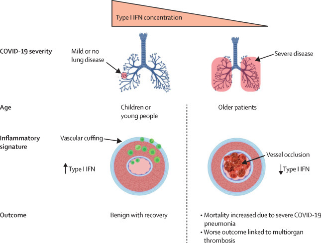
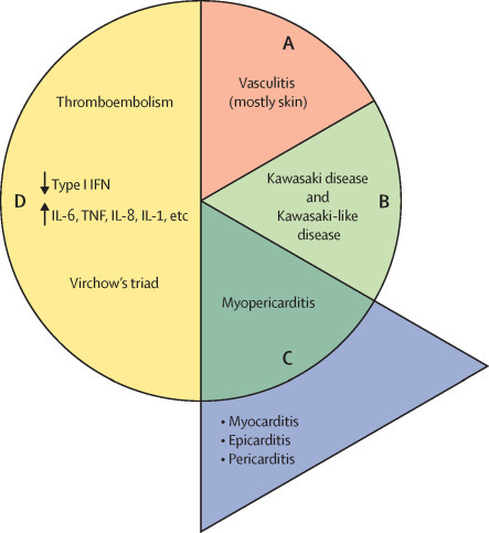
COVID-19 has been occasionally linked to histologically confirmed cutaneous vasculitis and a Kawasaki-like vasculitis, with these entities generally having minimal or no lung involvement and a good prognosis. Unlike these vasculitis types, patients with severe COVID-19 pneumonia can develop cutaneous vasculitis-like lesions and systemic arterial and venous thromboemboli, including cryptogenic strokes and other vasculopathy features. Proposed underlying mechanisms for these severe manifestations have encompassed immune dysregulation, including an anti-phospholipid syndrome-like state, complement activation, viral dissemination with direct systemic endothelial infection, viral RNAaemia with immunothrombosis, clotting pathway activation mediated by hypoxaemia, and immobility. In this Viewpoint, we highlight how imaging and post-mortem findings from patients with COVID-19 indicate a novel thrombosis in the pulmonary venous territory distal to the alveolar capillary bed, a territory that normally acts as a clot filtration system, which might represent an unappreciated nidus for systemic microembolism. Additionally, we suggest that this mechanism represents a novel vasculitis mimic related to COVID-19 that might lead to cryptogenic strokes across multivessel territories, acute kidney injury with haematuria, a skin vasculitis mimic, intestinal ischaemia, and other organ ischaemic manifestations. This finding is supported by pathological reports of extensive pulmonary venular thrombosis and peripheral organ thrombosis with pauci-immune cellular infiltrates. Therefore, severe COVID-19 pneumonia with extensive pulmonary intravascular coagulopathy might help to explain the numerous systemic complications of COVID-19, in which the demonstration of direct organ infection has not adequately explained the pathology.


Figures

Figure 1
Figure 1
Patterns of disease leading to vasculitis and vasculitis mimics Two distinct mechanisms underscore the cutaneous disease seen in COVID-19. For mild disease in younger patients, a transient vasculitis limited to the skin might occur in the context of robust immune responses to COVID-19, including vigorous type I IFN responses and mild lung disease. The basis for Kawasaki-related vasculitis, in general or in COVID-19, is poorly defined; however, it typically occurs in individuals with minimal or no lung infection. For severe COVID-19 pneumonia with respiratory failure, cutaneous vasculitis is linked to small vessel thromboembolic disease. This pattern could be partly due to thrombosis of small and large vessels in situ due to severe illness, hypoxaemia, and RNAaemia in some severe cases. The pattern might also be linked to diffuse embolism from the pulmonary venular, left heart, and arterial emboli dislodgement from thrombi. Cutaneous disease in patients with severe COVID-19 might be linked to type I IFN disablement and elevations in multiple proinflammatory cytokines. IFN=interferon.
Figure 2
Figure 2
Types of vasculitis disease A) Uncommon and mild cutaneous vasculitis is linked to robust immune responses, typically occurs in otherwise healthy patients, and involves skin only, with a predilection for the toes. This cutaneous vasculitis occurs in the context of robust type I IFN production and intact anti-SARS-CoV-2 immunity. B) Kawasaki disease can occur in patients with non-severe infection, presenting with classically described coronary aneurysms but with a pattern of skin involvement distinct to that in isolated mild cutaneous vasculitis. Kawasaki-like disease, seen in older children and young adults with non-severe infection, is associated with prominent cardiac muscle involvement and absence of aneurysms. C) Predominant myocarditis might occur as part of the Kawasaki phenotype and be the dominant clinical picture, and cases with cardiac inflammation might occur without any detectable aneurysm formation (blue panel). D) The vasculitis mimics seen in severe COVID-19 pneumonia, associated with cardiac disease, are likely to be linked to multifaceted mechanisms consequent to severe pulmonary territory immunothrombosis, hypoxaemia, pulmonary hypertension, pulmonary venular territory embolism, arterial thromboembolism, and systemic venous thromboembolism. Decreased type I IFN responses and subsequent activation of proinflammatory cytokines (IL-1, IL-6, IL-8, TNF, etc) and chemokines result in immunothrombosis. IFN=interferon. IL=interleukin. SARS-CoV-2=severe acute respiratory syndrome coronavirus 2. TNF=tumour necrosis factor.
Figure 3
Figure 3
Sources of thromboembolic material for vasculitis mimics in COVID-19 SARS-CoV-2 infection might lead to a vasculitis mimic by two major mechanisms. First, local pulmonary intravascular coagulopathy might lead to embolisation originating in the pulmonary venous vasculature location, given that it is a well established site of thrombosis in COVID-19 and readily fulfils Virchow's triad outside of pulmonary venular walls, inflammation within pulmonary venule walls, change in flow due to decreased capillary drainage, and probable changes within the vessel lumen due to clotting cascade factors and immunothrombotic SARS-CoV-2 viral RNA. Given that the pulmonary vein system has thinner walls than the pulmonary arterial system, the vein system could also favour the effect of exogenous alveolar inflammation spread to this territory with extensive venular immunothrombosis. Second, a systemic hypercoagulopathy might exist due to dissemination of viral RNA that triggers immunothrombosis outside of the pulmonary circulation in the arterial and venous trees. Such a scenario might lead to embolisation originating from the heart or the arterial tree itself. SARS-CoV-2=severe acute respiratory syndrome coronavirus 2.
Figure 4
Figure 4
Basis for pulmonary venule microthrombosis with embolism A) Pulmonary venule thrombosis occurs due to severe and proven SARS-CoV-2 centric alveolitis. Inflammatory mediators and inflammatory cells outside of the thin-walled pulmonary venular circulation; immune cell infiltration of venular walls; activated immune cells within venules; and release of procoagulation tissue factor and other procoagulants, and proinflammatory mediators trigger venular thrombosis. Activation of the fibrinolytic pathway, as evidenced by increased D-dimers and respiratory distress, lung tissue movement, and potential impact of barotrauma in ventilated patients with severe COVID-19 all contribute to the proposed systemic emboli derived from the pulmonary vein territory. Systemic microemboli that originate beyond the pulmonary capillary filtration network access the circulation. Pulmonary venule thrombosis is evident histologically, and CT scanning of the chest frequently shows pre-obstruction vascular dilatation. B) The main difference between an immunothrombotic clot and a classical clot is that the classical type is predominately composed of fibrin and platelets, rather than immune cells. SARS-CoV-2=severe acute respiratory syndrome coronavirus 2.

References

    1. Al-Ani F, Chehade S, Lazo-Langner A. Thrombosis risk associated with COVID-19 infection. A scoping review. Thromb Res. 2020;192:152–160. - PMC - PubMed
    1. Fox SE, Akmatbekov A, Harbert JL, Li G, Quincy Brown J, Vander Heide RS. Pulmonary and cardiac pathology in African American patients with COVID-19: an autopsy series from New Orleans. Lancet Respir Med. 2020;8:681–686. - PMC - PubMed
    1. O'Sullivan JM, Gonagle DM, Ward SE, Preston RJS, O'Donnell JS. Endothelial cells orchestrate COVID-19 coagulopathy. Lancet Haematol. 2020;7:e553–e555. - PMC - PubMed
    1. McGonagle D, O'Donnell JS, Sharif K, Emery P, Bridgewood C. Immune mechanisms of pulmonary intravascular coagulopathy in COVID-19 pneumonia. Lancet Rheumatol. 2020;2:e437–e445. - PMC - PubMed
    1. McGonagle D, Plein S, O'Donnell JS, Sharif K, Bridgewood C. Increased cardiovascular mortality in African Americans with COVID-19. Lancet Respir Med. 2020;8:649–651. - PMC - PubMed

LinkOut - more resources