. 2021 Jan 19;2(1):100185.
doi: 10.1016/j.xcrm.2020.100185.

Stronger induction of trained immunity by mucosal BCG or MTBVAC vaccination compared to standard intradermal vaccination

Affiliations

Stronger induction of trained immunity by mucosal BCG or MTBVAC vaccination compared to standard intradermal vaccination

Michel P M Vierboom et al. Cell Rep Med. .

Abstract

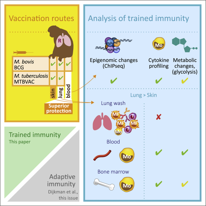
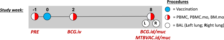
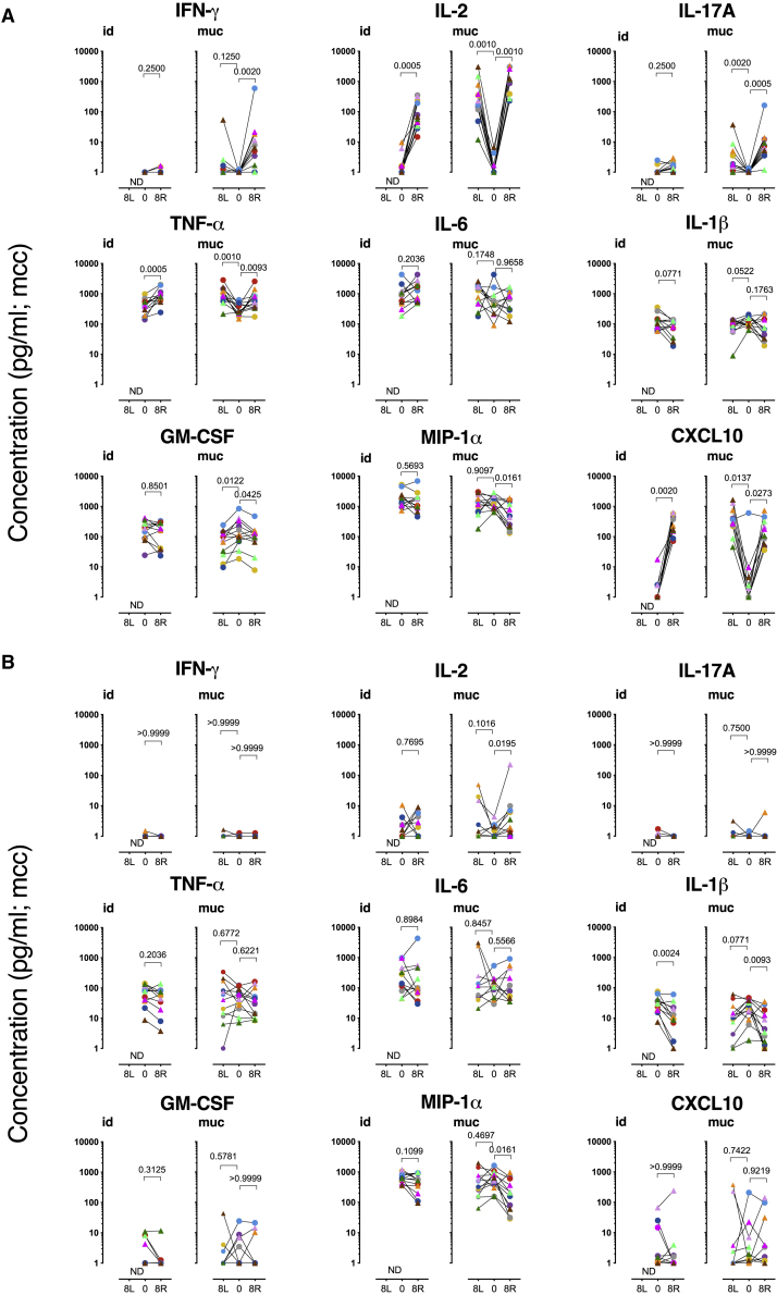
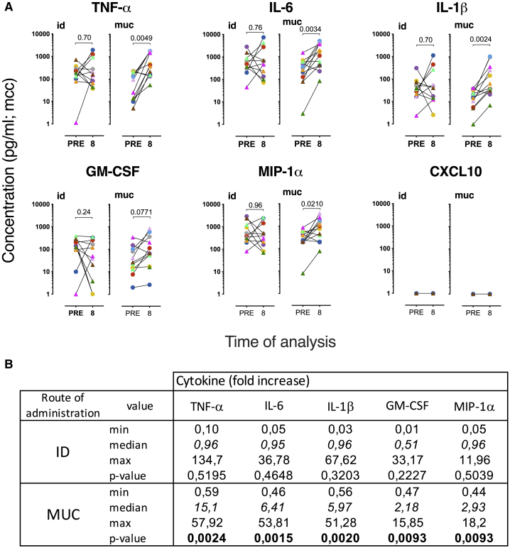
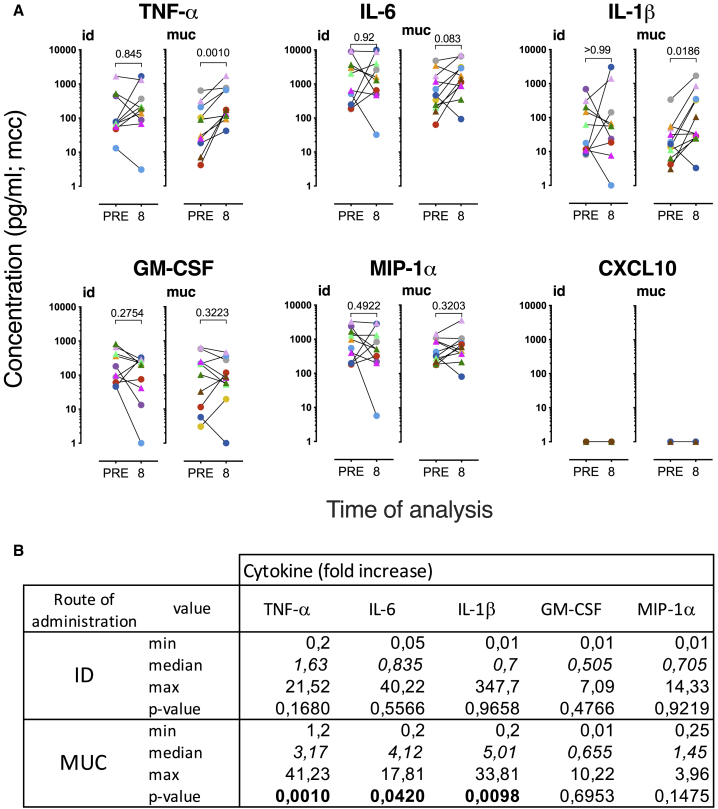
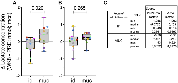
BCG vaccination can strengthen protection against pathogens through the induction of epigenetic and metabolic reprogramming of innate immune cells, a process called trained immunity. We and others recently demonstrated that mucosal or intravenous BCG better protects rhesus macaques from Mycobacterium tuberculosis infection and TB disease than standard intradermal vaccination, correlating with local adaptive immune signatures. In line with prior mouse data, here, we show in rhesus macaques that intravenous BCG enhances innate cytokine production associated with changes in H3K27 acetylation typical of trained immunity. Alternative delivery of BCG does not alter the cytokine production of unfractionated bronchial lavage cells. However, mucosal but not intradermal vaccination, either with BCG or the M. tuberculosis-derived candidate MTBVAC, enhances innate cytokine production by blood- and bone marrow-derived monocytes associated with metabolic rewiring, typical of trained immunity. These results provide support to strategies for improving TB vaccination and, more broadly, modulating innate immunity via mucosal surfaces.

Keywords: BCG; MTBVAC; immunotherapy; innate immunity; non-human primates; trained immunity; tuberculosis; vaccination; vaccine development.


Conflict of interest statement

E.R., E.P., N.A., and C.M. are co-inventors on a patent on a tuberculosis vaccine held by the University of Zaragoza and Biofabri. E.R. and E.P. are employees of Biofabri. J.T. is an employee of the TuBerculosis Vaccine Initiative and an advisor to Biofabri. M.G.N. holds a patent on the inhibition of trained immunity with nanobiologics, and a patent on the stimulation of trained immunity with nanobiologics. M.G.N. is also a scientific founder of Trained Therapeutix and Discovery (TTxD). The remaining authors of the Radboud UMC and the authors from the BPRC declare no competing interests.

Figures

None
Graphical abstract
Figure 1
Figure 1
Schematic representation of the timeline of vaccination in the different vaccination strategies The BCG.iv group (n = 3) was analyzed at weeks −1 and 2. The groups that were immunized with BCG.id (n = 6), BCG.muc (n = 6), MTBVAC.id (n = 6), and MTBVAC.muc (n = 6) were analyzed at weeks −1 and 8. Only from mucosally vaccinated animals BAL was collected from the right lung as well as the left lung. See also Table S1.
Figure 2
Figure 2
Mucosal vaccination establishes a unique local adaptive signature defined by polyfunctional Th17 cells (A–C) Individual IFN-γ ELISpot responses after in vitro recall stimulation of PBMCs against PPD (A) before vaccination or (B) 8 weeks after vaccination or with (C) ESAT6-CFP10 8 weeks after vaccination. Horizontal lines in bars indicate group medians; n = 6 animals/group. ANOVA adjusted for multiple comparisons; Dunn’s multiple comparison test. (D) The percentages of polyfunctional IL-17A+ PPD-specific CD4+ T cells (also producing IFN-γ, TNF-α, and IL-2) were determined by flow cytometric analysis of BAL cells before (PRE) and 8 weeks after intradermal (.id) or mucosal (.muc) vaccination. Cells were typically collected from the lower right lung lobe (R); targeted for vaccination) and for mucosally vaccinated animals also from the left lung (L) to establish dissemination of the immune response. The data are presented as medium control corrected (cc); n = 6 animals/group; Wilcoxon matched-pairs signed rank test. ULD, upper limit of detection; LLD, lower limit of detection. See also Figure S7.
Figure 3
Figure 3
Trained immunity after intravenous BCG vaccination (A and B) Freshly isolated BAL cells (from the right lung) and monocytes from blood (PBMC.mo) and bone marrow (BM.mo) were stimulated for 24 h with (A) Mtb whole-cell lysate (25 μg/mL) or (B) LPS (0.1 μg/mL) before and 2 weeks after intravenous vaccination with BCG. The data are presented as medium control corrected. (C) Lactate production as an indicator of metabolic rewiring was measured in 24-h supernatant from LPS-stimulated monocytes purified from blood (PBMC.mo) or BM (BM.mo). See also Figure S1. (D) Heatmap of H3K27ac reads (red) over BCG-specific peaks. The intensity over the center of the peak ± 12 kb is depicted. (E) The top GO pathways associated with the nearest genes to dynamic H3K27ac, with adjusted p values. (F) H3K27ac dynamics at interferon regulatory factor 3 (IRF3) and syndecan 2 (SDC2) locus for PBMC.mo pre- and post-vaccination. The complete list of genes and values can be found at GEO: GSE159046.
Figure 4
Figure 4
Cytokine production by BAL cells Freshly isolated BAL cells were stimulated (A) with Mtb whole cell lysate or (B) LPS, before (PRE) and 8 weeks after intradermal (.id) or mucosal (.muc) vaccination (with either BCG, in circles, or MTBVAC, in triangles). Cells were typically collected from the lower right lung lobe (R), the lobe targeted by vaccination, and for mucosally vaccinated animals also from the left lung (L), to establish dissemination of the immune response. The data are presented as medium control corrected (mcc); n = 12 animals/group; Wilcoxon matched-pairs signed rank test. p values are indicated at the top of each graph. See also Figure S2.
Figure 5
Figure 5
Trained immunity after mucosal vaccination in peripheral blood monocytes Cytokine production was measured after 24 h of stimulation with LPS (0.1 μg/mL). (A) Paired display of cytokine levels before and 8 weeks after vaccination. The data are presented as medium control corrected (mcc; BCG in circles; MTBVAC in triangles). Statistical significance was calculated by Wilcoxon’s non-parametric paired analysis test (n = 12 animals/group; PRE versus 8 weeks post-vaccination). p values are indicated at the top of each graph. (B) Median fold increase in cytokine production is indicated in italics; significant p < 0.05 is indicated in bold; n = 12 animals/group; Wilcoxon signed rank test. See also Figures S1, S3, S4, S5, and S6.
Figure 6
Figure 6
Trained immunity after mucosal vaccination in BM monocytes Cytokine production was measured after 24 h of stimulation with LPS (0.1 μg/mL). (A) Paired display of cytokine levels before and 8 weeks after vaccination. The data are presented as medium control corrected (mcc; BCG in circles; MTBVAC in triangles). Statistical significance was calculated by Wilcoxon’s non-parametric paired analysis test (n = 12 animals/group; PRE versus 8 weeks post-vaccination). p values are indicated at the top of each graph. (B) Median fold increase in cytokine production is indicated in italics; significant p < 0.05 is indicated in bold; n = 12 animals/group; Wilcoxon signed rank test. See also Figures S1, S4, and S6.
Figure 7
Figure 7
Increased lactate production after mucosal vaccination (A and B) Lactate production as an indicator of metabolic rewiring was measured in 24-h supernatant from LPS-stimulated monocytes purified (A) from peripheral blood, or (B) BM. Statistical significance between group median values was calculated by Mann-Whitney rank testing and indicated at the top. The box extends from the 25th to 75th percentiles while the whiskers go down to the smallest value and up to the largest. (C) Median fold increase in lactate production is indicated in italics; significant p < 0.05 is indicated in bold; n = 12 animals/group; Wilcoxon signed rank test. See also Figure S6.

References

    1. Kaufmann S.H.E. Immunology’s Coming of Age. Front. Immunol. 2019;10:684. - PMC - PubMed
    1. Netea M.G., Joosten L.A., Latz E., Mills K.H., Natoli G., Stunnenberg H.G., O’Neill L.A., Xavier R.J. Trained immunity: A program of innate immune memory in health and disease. Science. 2016;352:aaf1098. - PMC - PubMed
    1. Kleinnijenhuis J., Quintin J., Preijers F., Joosten L.A., Ifrim D.C., Saeed S., Jacobs C., van Loenhout J., de Jong D., Stunnenberg H.G. Bacille Calmette-Guerin induces NOD2-dependent nonspecific protection from reinfection via epigenetic reprogramming of monocytes. Proc. Natl. Acad. Sci. USA. 2012;109:17537–17542. - PMC - PubMed
    1. Cheng S.C., Quintin J., Cramer R.A., Shepardson K.M., Saeed S., Kumar V., Giamarellos-Bourboulis E.J., Martens J.H., Rao N.A., Aghajanirefah A. mTOR- and HIF-1α-mediated aerobic glycolysis as metabolic basis for trained immunity. Science. 2014;345:1250684. - PMC - PubMed
    1. Quintin J., Saeed S., Martens J.H.A., Giamarellos-Bourboulis E.J., Ifrim D.C., Logie C., Jacobs L., Jansen T., Kullberg B.J., Wijmenga C. Candida albicans infection affords protection against reinfection via functional reprogramming of monocytes. Cell Host Microbe. 2012;12:223–232. - PMC - PubMed

Publication types

MeSH terms

LinkOut - more resources