. 2021 Apr 9;2(4):411-422.e5.
doi: 10.1016/j.medj.2021.01.001. Epub 2021 Jan 16.

Cell-free DNA tissues of origin by methylation profiling reveals significant cell, tissue, and organ-specific injury related to COVID-19 severity

Affiliations

Cell-free DNA tissues of origin by methylation profiling reveals significant cell, tissue, and organ-specific injury related to COVID-19 severity

Alexandre Pellan Cheng et al. Med. .

Abstract

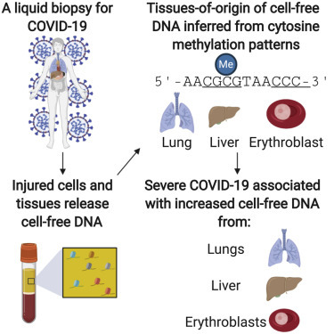
Background: Coronavirus disease 2019 (COVID-19) primarily affects the lungs, but evidence of systemic disease with multi-organ involvement is emerging. Here, we developed a blood test to broadly quantify cell-, tissue-, and organ-specific injury due to COVID-19.

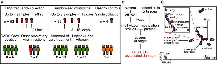
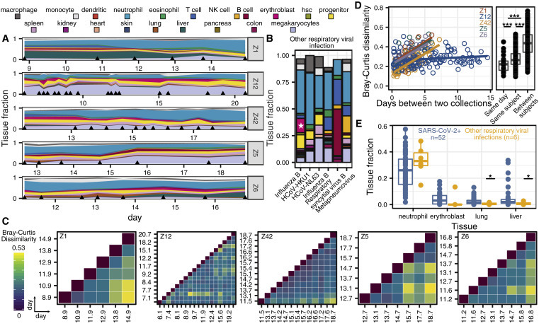
Methods: Our test leverages genome-wide methylation profiling of circulating cell-free DNA in plasma. We assessed the utility of this test to identify subjects with severe disease in two independent, longitudinal cohorts of hospitalized patients. Cell-free DNA profiling was performed on 104 plasma samples from 33 COVID-19 patients and compared to samples from patients with other viral infections and healthy controls.

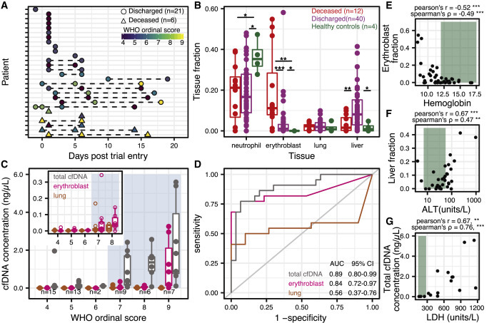
Findings: We found evidence of injury to the lung and liver and involvement of red blood cell progenitors associated with severe COVID-19. The concentration of cell-free DNA correlated with the World Health Organization (WHO) ordinal scale for disease progression and was significantly increased in patients requiring intubation.

Conclusions: This study points to the utility of cell-free DNA as an analyte to monitor and study COVID-19.

Funding: This work was supported by NIH grants 1DP2AI138242 (to I.D.V.), R01AI146165 (to I.D.V., M.P.C., F.M.M., and J.R.), 1R01AI151059 (to I.D.V.), K08-CA230156 (to W.G.), and R33-AI129455 to C.Y.C., a Synergy award from the Rainin Foundation (to I.D.V.), a SARS-CoV-2 seed grant at Cornell (to I.D.V.), a National Sciences and Engineering Research Council of Canada fellowship PGS-D3 (to A.P.C.), and a Burroughs-Wellcome CAMS Award (to W.G.). D.C.V. is supported by a Fonds de la Recherche en Sante du Quebec Clinical Research Scholar Junior 2 award. C.Y.C. is supported by the California Initiative to Advance Precision Medicine, and the Charles and Helen Schwab Foundation.

Keywords: COVID-19; SARS-CoV-2; cell-free DNA; disease severity; liquid biopsy; methylation; tissue injury.


Conflict of interest statement

A.P.C., M.P.C., W.G., C.Y.C., D.C.V., and I.D.V. are inventors on a patent application submitted by Cornell University Center for Technology Licensing.

Figures

None
Graphical abstract
Figure 1
Figure 1
Study design (A) Two independent cohorts were used in our study. First, a high-frequency collection cohort with 5 SARS-CoV-2 patients (n = 52 samples) and 6 SARS-CoV-2-negative, RNA virus-positive patients (n = 6 samples). Second, a randomized controlled trial of 28 SARS-CoV-2 patients with plasma at serial time points (n = 52 samples). Four healthy individuals volunteered plasma for cell-free DNA analysis. (B) Experimental workflow. cfDNA is extracted from plasma, and whole-genome bisulfite sequencing is performed. In parallel, methylation profiles of cell and tissue genomes are obtained from publicly available databases. cfDNA methylation profiles are compared to those of cell and tissue references to infer relative contributions of tissues to the cfDNA mixtures. (C) UMAP of differentially methylated regions for isolated cell and tissue types used as a reference.
Figure 2
Figure 2
High-Frequency sample collection cohort at UCSF (A and B) Patient-specific relative tissue contributions for SARS-CoV-2 patients (A) and other RNA virus infection patients (B). Triangles (A) indicate sampling times and the star (B) represents the erythroblast fraction of an influenza B patient who was being treated for recurrent stage IV diffuse large B cell lymphoma. (C) Heatmaps of Bray-Curtis dissimilarity. (D) Scatterplot of patient-specific Bray-Curtis dissimilarity (left) and boxplot of Bray-Curtis dissimilarity between cfDNA tissue proportions from samples collected from either the same day (within 24 h), the same person (but not within 24 h), or from all of the patients (right). (E) Comparison of tissue fraction of 4 cell and tissue types (neutrophil, erythroblast, lung, and liver) between SARS-CoV-2-positive patients and other RNA virus-positive patients. ∗p < 0.05; ∗∗p < 0.01; ∗∗∗p < 0.001 (p values calculated using a Wilcoxon test)
Figure 3
Figure 3
Randomized controlled trial cohort from MUHC (A) Patient sample collection map by day of enrollment in the study. (B) Relative proportion of cfDNA derived from 4 cell and tissue types (neutrophil, erythroblast, lung, liver) by hospitalization status (p values calculated using a Wilcoxon test). (C) Absolute cfDNA concentrations compared to the WHO ordinal scale for COVID progression. Blue shading indicates ordinal scores requiring admittance to the intensive care unit (ICU). (D) Receiver operating characteristic analysis of the performance of absolute cfDNA concentration of different tissues (lung, erythroblast, and total) in distinguishing patients presenting with ordinal scales from 4 to 6 (hospitalized) and 7 to 9 (hospitalized in the ICU). (E–G) Scatterplot comparisons between relative proportions of erythroblast cfDNA fraction and hemoglobin (E), liver cfDNA fraction and alanine aminotransferase (ALT) (F), and total cfDNA concentration and lactase dehydrogenase (LDH) (H). Green shading indicates normal levels. ∗p < 0.05; ∗∗p < 0.01; ∗∗∗p < 0.001.

Update of

References

    1. Fox S.E., Akmatbekov A., Harbert J.L., Li G., Quincy Brown J., Vander Heide R.S. Pulmonary and cardiac pathology in African American patients with COVID-19: an autopsy series from New Orleans. Lancet Respir. Med. 2020;8:681–686. - PMC - PubMed
    1. Puelles V.G., Lütgehetmann M., Lindenmeyer M.T., Sperhake J.P., Wong M.N., Allweiss L., Chilla S., Heinemann A., Wanner N., Liu S., et al. Multiorgan and Renal Tropism of SARS-CoV-2. N. Engl. J. Med. 2020;383:590–592. - PMC - PubMed
    1. Gupta A., Madhavan M.V., Sehgal K., Nair N., Mahajan S., Sehrawat T.S., Bikdeli B., Ahluwalia N., Ausiello J.C., Wan E.Y., et al. Extrapulmonary manifestations of COVID-19. Nat. Med. 2020;26:1017–1032. - PMC - PubMed
    1. Varga Z., Flammer A.J., Steiger P., Haberecker M., Andermatt R., Zinkernagel A.S., Mehra M.R., Schuepbach R.A., Ruschitzka F., Moch H. Endothelial cell infection and endotheliitis in COVID-19. Lancet. 2020;395:1417–1418. - PMC - PubMed
    1. Julien P., Julien G., Morgan C., et al. Pulmonary Embolism in COVID-19 Patients: Awareness of an Increased Prevalence. Circulation. 2020;142:184–186. - PubMed

Publication types

Substances