. 2021 Mar 15;60(12):6480-6487.
doi: 10.1002/anie.202012046. Epub 2021 Feb 12.

Histone H4 Tails in Nucleosomes: a Fuzzy Interaction with DNA

Affiliations

Histone H4 Tails in Nucleosomes: a Fuzzy Interaction with DNA

Sevastyan O Rabdano et al. Angew Chem Int Ed Engl. .

Abstract

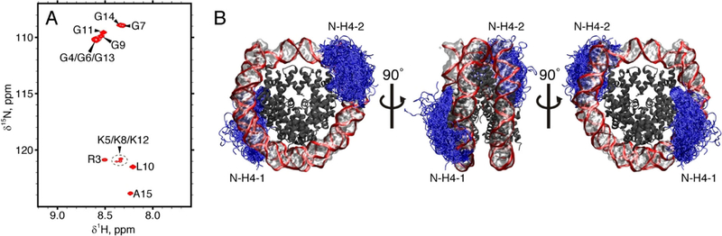
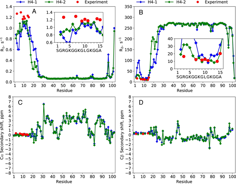
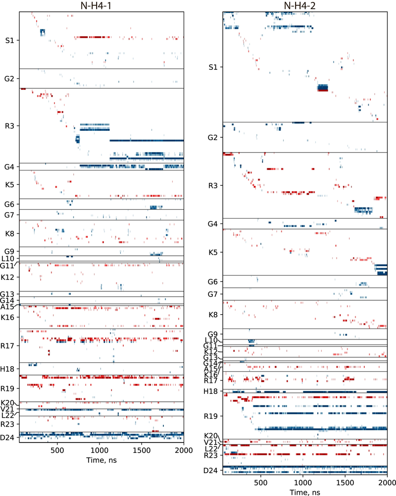
The interaction of positively charged N-terminal histone tails with nucleosomal DNA plays an important role in chromatin assembly and regulation, modulating their susceptibility to post-translational modifications and recognition by chromatin-binding proteins. Here, we report residue-specific 15 N NMR relaxation rates for histone H4 tails in reconstituted nucleosomes. These data indicate that H4 tails are strongly dynamically disordered, albeit with reduced conformational flexibility compared to a free peptide with the same sequence. Remarkably, the NMR observables were successfully reproduced in a 2-μs MD trajectory of the nucleosome. This is an important step toward resolving an apparent inconsistency where prior simulations were generally at odds with experimental evidence on conformational dynamics of histone tails. Our findings indicate that histone H4 tails engage in a fuzzy interaction with nucleosomal DNA, underpinned by a variable pattern of short-lived salt bridges and hydrogen bonds, which persists at low ionic strength (0-100 mM NaCl).

Keywords: NMR spectroscopy; fuzzy protein-DNA interactions; histone tails; molecular dynamics; nucleosome.


Conflict of interest statement

Conflict of interest

The authors declare no conflict of interest.

Supporting information and the ORCID identification number(s) for the author(s) of this article can be found under: https://doi.org/10.1002/anie.202012046.

Figures

Figure 1.
Figure 1.
(A) 1H,15N-HSQC spectrum of mononucleosomes containing 15N-labeled H4. (B) Dynamics of N-H4 tails (pictured as blue tubes) in mononucleosome according to the MD simulation data. To generate this plot, a 2-μs trajectory of NCP in TIP4P-D water was sampled with the step of 10 ns; the extracted frames were overlaid onto the reference structure 3LZ0. The DNA backbone is shown as a red band, bases and sugars are shown as a semi-transparent surface, the bodies of histone proteins are dark grey (with tails of H3, H2A, and H2B histones not shown). The visible asymmetry in spatial distribution of the N-H4–1 and N-H4–2 tails reflects a limited convergence of the simulation.
Figure 2.
Figure 2.
(A,B) 15N relaxation rate constants R1, R2 and (C,D) 13Cα/β secondary chemical shifts δsec for H4 histone proteins in NCP. Experimental values are shown with red circles, the results of MD-based calculations—with blue and green diamonds (first and second copy of H4, as enumerated in the structure 3LZ0). The MD trajectory is that of NCP in TIP4P-D water, with a net length of 2 μs. Additionally, 1HN and 15N δsec data are summarized in Figure S5.
Figure 3.
Figure 3.
Traces of the interactions between N-terminal tails of H4 histones (two copies in the NCP, residues 1–24) and the remaining part of the NCP. Each horizontal band represents a specific interaction involving one of the tail residues (indicated on the y-axis). For a given residue, interactions are sorted according to the time of the first appearance; the ordered list that identifies all interactions can be found in Table S2. Hydrogen bonds and salt bridges are coded blue and red, respectively. The definition of salt bridges between DNA phosphate group and arginine or lysine side chains is illustrated in Figure S6.

Similar articles

Cited by

References

    1. Boublik M, Bradbury EM, Crane-Robinson C, Johns EW, Eur. J. Biochem 1970, 17, 151–159; - PubMed
    2. Lilley DMJ, Howarth OW, Clark VM, Pardon JF, Richards BM, FEBS Lett 1976, 62, 7–10. - PubMed
    1. Drozdetskiy A, Cole C, Procter J, Barton GJ, Nucleic Acids Res 2015, 43, W389–W394. - PMC - PubMed
    1. Cary PD, Crane-Robinson C, Bradbury EM, Dixon GH, Eur. J. Biochem 1982, 127, 137–143. - PubMed
    1. Weintraub H, Vanlente F, Proc. Natl. Acad. Sci. USA 1974, 71, 4249–4253; - PMC - PubMed
    2. Walker IO, Biochemistry 1984, 23, 5622–5628; - PubMed
    3. Hilliard PR, Smith RM, Rill RL, J. Biol. Chem 1986, 261, 5992–5998; - PubMed
    4. Stefanovsky VY, Dimitrov SI, Russanova VR, Angelov D, Pashev IG, Nucleic Acids Res 1989, 17, 10069–10081. - PMC - PubMed
    1. Kale S, Goncearenco A, Markov Y, Landsman D, Panchenko AR, Curr. Opin. Struct. Biol 2019, 56, 164–170. - PMC - PubMed

Publication types

LinkOut - more resources