Comparative Study
. 2021 Apr;13(2):474-483.
doi: 10.1111/os.12856. Epub 2021 Jan 31.

Anterior Controllable Antedisplacement and Fusion (ACAF) vs Posterior Laminoplasty for Multilevel Severe Cervical Ossification of the Posterior Longitudinal Ligament: Retrospective Study Based on a Two-Year Follow-up

Affiliations
Comparative Study

Anterior Controllable Antedisplacement and Fusion (ACAF) vs Posterior Laminoplasty for Multilevel Severe Cervical Ossification of the Posterior Longitudinal Ligament: Retrospective Study Based on a Two-Year Follow-up

Qing-Jie Kong et al. Orthop Surg. 2021 Apr.

Abstract

Objectives: To compare the clinical outcomes of anterior controllable antedisplacement fusion (ACAF), a new surgical technique, with laminoplasty for the treatment of multilevel severe cervical ossification of the posterior longitudinal ligament (OPLL) based on a 2-year follow-up.

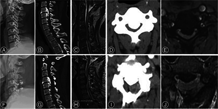
Methods: Clinical data of 53 patients (21 by ACAF and 32 by laminoplasty) who have accepted surgery for treatment of cervical myelopathy caused by multilevel severe OPLL (occupying rate ≥ 50%) from March 2015 to March 2017 were retrospectively reviewed and compared between ACAF group and laminoplasty group. Operative time, blood loss, and complications of the two groups were recorded. Radiographic parameters were evaluated pre- and postoperatively: cervical lordosis on X-ray, space available for the cord (SAC) and the occupying ratio (OR) on computed tomography (CT), and the anteroposterior (AP) diameter of the spinal cord at the narrowest level and the spinal cord curvature on magnetic resonance imaging (MRI). Japanese Orthopaedic Association (JOA) scoring was used to evaluate neurologic recovery. Statistical analysis was conducted to analyze the differences between two groups. The Mann-Whitney U test and chi square test were used to compare categorical variables. unpaired t test was used to compare continuous data.

Results: All patients were followed up for at least 24 months. The operative time was longer in ACAF group (286.5 vs 178.2 min, P < 0.05). The blood loss showed no significant difference (291.6 vs 318.3 mL, P > 0.05). Less complications were observed in ACAF group than in laminoplasty group (one case [4.7%] of C5 palsy and one case [4.7%] of cerebrospinal fluid [CSF] leakage in ACAF group; four cases [12.5%] of C5 palsy, two cases [6.3%] of CSF leakage, and four cases [12.5%] of axial symptoms in laminoplasty group). The mean JOA score at last follow-up (14.6 vs 12.8, P < 0.05) and the improvement rate (IR) (63.8% vs 47.8%, P < 0.05) in ACAF group were superior to those in laminoplasty group significantly. The postoperative OR (16.7% vs 40.9%, P < 0.05), SAC (150.8 vs 110.5 mm2 , P < 0.05), AP spinal cord diameter (5.5 vs 4.2 mm, P < 0.05), and cervical lordosis (12.7° vs 4.7°, P < 0.05) were improved more considerably in ACAF group, with significant differences between two groups. Notably, the spinal cord on MRI showed a better curvature in ACAF group.

Conclusions: This study showed that ACAF is considered superior to laminoplasty for the treatment of multilevel severe OPLL as anterior direct decompression and better curvature of the spinal cord led to satisfactory neurologic outcomes and low complication rate.

Keywords: Anterior controllable antedisplacement and fusion (ACAF); Complication; Laminoplasty; Myelopathy; Ossification of the posterior longitudinal ligament (OPLL).


Figures

Fig. 1
Fig. 1
Schematic diagram for ACAF surgery. (A) the ossification of posterior longitudinal ligament at C4–C6; (B) resection of C3/4, C4/5, C5/6, and C6/7 intervertebral discs and remove the anterior bone of C4–C6 vertebrae; (C) grooving at the operator's opposite side, partially grooving at the operator's side; (D) installing cages, screws, and titanium plate and complete grooving at the operator's side; (E) sagittal view after installing internal fixation; (F) hoisting C4–C6 vertebrae.
Fig. 2
Fig. 2
ACAF surgery. (A, G) Preoperative CT and MRI sagittal scan demonstrated spinal stenosis from C3 to C5, and severe OPLL from C4 to C5. The red dash line square showed the vertebral‐OPLL complex (VOC) levels. (B) The red dash line square showed the cut part of the anterior bone with the thickness according to ossified ligament. (C) Postoperative CT coronal reconstruction demonstrated the bilateral osteotomies conducted at the inner border of uncovertebral joints. (D) The red dash line square showed the cut part of the anterior bone with the thickness according to ossified ligament. And the red line indicated the position of grooves on both sides. (E) The red arrows showed the position of grooves on both sides. After tightening the screw in the middle vertebrae in a hoisting manner, the VOC moved forward. Then the space available for the cord were restored. (F‐H) Postoperative CT and MRI sagittal scan demonstrated satisfactory antedisplacement of VOC from C4 to C5. Cervical lordosis and space available for the cord were restored.
Fig. 3
Fig. 3
Radiological measurements for both groups. (A) the occupation rate (OR), (a) proper anteroposterior diameter of the spinal canal, (b) thickness of the ossification at the level of the greatest canal narrowing. The OR is defined as (b) divided by (a). (B) space available for the cord (SAC). (C) anteroposterior (AP) diameter of the spinal cord. (D) cervical lordosis. Left, ACAF group (A1, B1, C1, and D1, respectively); right, Laminoplasty group (A2, B2, C2, and D2, respectively).
Fig. 4
Fig. 4
Five types of spinal cord curvature after operation. (A) Type I, lordosis. (B) Type II, straight with no shifting. (C) Type III, straight with shifting. (D) Type IV, sigmoid. (E) Type V, kyphosis.
Fig. 5
Fig. 5
A 55‐year‐old woman undergoing ACAF. (A) Lateral X‐ray showing the cervical lordosis was 5°. (B) sagittal CT of the cervical spine showing spinal stenosis from C3 to C5, and severe OPLL from C4 to C5. (C) Sagittal MRI showing spinal cord compression from C3 to C5, especially from C4 to C5. (D) Cross‐sectional CT showing the occupying rate of 73.2%. (E) The morphology of the spinal cord on cross‐sectional MRI was crescent and diameter of spinal cord was 2.5 mm. (F) Lateral X‐ray showing a surgery of ACAF from C3 to C6 and the cervical lordosis was 15°. (G) Sagittal CT and (H) MRI demonstrating the spinal canal returning to normal volume and sufficient decompression and the type of spinal cord curvature was Type I lordosis. (I) Cross‐sectional CT showing the occupying rate of 4.5%. (J) The morphology of the spinal cord on cross‐sectional MRI was cylinder and diameter of spinal cord was 5.8 mm.
Fig. 6
Fig. 6
A 57‐year‐old man undergoing laminoplasty. (A) Lateral X‐ray showing the cervical lordosis was 5°. (B) sagittal CT of the cervical spine showing spinal stenosis from C4 to C6, and severe OPLL from C4 to C5. (C) Sagittal MRI showing spinal cord compression from C4 to C6, especially from C4 to C5. (D) Cross‐sectional CT showing the occupying rate of 70.5%. (E) The morphology of the spinal cord on cross‐sectional MRI was boomerang and diameter of spinal cord was 2.3 mm. (F) Lateral X‐ray showing a surgery of laminoplasty from C4 to C6 and the cervical lordosis was 6°. (G) Sagittal CT and (H) MRI demonstrating partial recovery of spinal canal and the type of spinal cord curvature was Type IV sigmoid. (I) Cross‐sectional CT showing the occupying rate of 44.0%. (J) The morphology of the spinal cord on cross‐sectional MRI was crescent and diameter of spinal cord was 3.6 mm.

References

    1. Abiola R, Rubery P, Mesfin A. Ossification of the posterior longitudinal ligament: etiology, diagnosis, and outcomes of nonoperative and operative management. Global Spine J, 2016, 6: 195–204. - PMC - PubMed
    1. Chen Y, Yang L, Liu Y, Yang H, Wang X, Chen D. Surgical results and prognostic factors of anterior cervical Corpectomy and fusion for ossification of the posterior longitudinal ligament. PLoS One, 2014, 9: e102008. - PMC - PubMed
    1. Edwards CC, Heller JG, Murakami H. Corpectomy versus Laminoplasty for multilevel cervical myelopathy. Spine (Phila Pa 1976), 2002, 27: 1168–1175. - PubMed
    1. Fujimori T, Iwasaki M, Okuda S, et al. Long‐term results of cervical myelopathy due to ossification of the posterior longitudinal ligament with an occupying ratio of 60% or more. Spine (Phila Pa 1976), 2014, 39: 58–67. - PubMed
    1. Nishida N, Kanchiku T, Kato Y, Imajo Y, Taguchi T. Cervical ossification of the posterior longitudinal ligament: factors affecting the effect of posterior decompression. J Am Paraplegia Soc, 2016, 40: 93–99. - PMC - PubMed

Publication types