

Cerebrospinal fluid flow in normal beagle dogs analyzed using magnetic resonance imaging
- PMID: 33522154
- PMCID: PMC7850791
- DOI: 10.4142/jvs.2021.22.e2
Cerebrospinal fluid flow in normal beagle dogs analyzed using magnetic resonance imaging
Abstract
Background: Diseases related to cerebrospinal fluid flow, such as hydrocephalus, syringomyelia, and Chiari malformation, are often found in small dogs. Although studies in human medicine have revealed a correlation with cerebrospinal fluid flow in these diseases by magnetic resonance imaging, there is little information and no standard data for normal dogs.
Objectives: The purpose of this study was to obtain cerebrospinal fluid flow velocity data from the cerebral aqueduct and subarachnoid space at the foramen magnum in healthy beagle dogs.
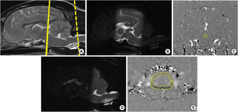
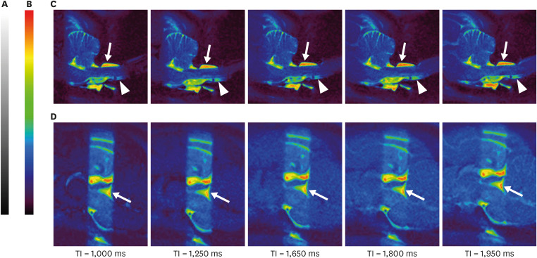
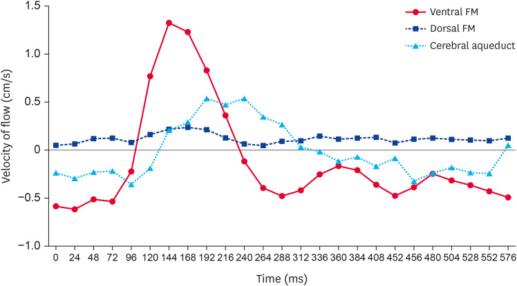
Methods: Six healthy beagle dogs were used in this experimental study. The dogs underwent phase-contrast and time-spatial labeling inversion pulse magnetic resonance imaging. Flow rate variations in the cerebrospinal fluid were observed using sagittal time-spatial labeling inversion pulse images. The pattern and velocity of cerebrospinal fluid flow were assessed using phase-contrast magnetic resonance imaging within the subarachnoid space at the foramen magnum level and the cerebral aqueduct.
Results: In the ventral aspect of the subarachnoid space and cerebral aqueduct, the cerebrospinal fluid was characterized by a bidirectional flow throughout the cardiac cycle. The mean ± SD peak velocities through the ventral and dorsal aspects of the subarachnoid space and the cerebral aqueduct were 1.39 ± 0.13, 0.32 ± 0.12, and 0.76 ± 0.43 cm/s, respectively.
Conclusions: Noninvasive visualization of cerebrospinal fluid flow movement with magnetic resonance imaging was feasible, and a reference dataset of cerebrospinal fluid flow peak velocities was obtained through the cervical subarachnoid space and cerebral aqueduct in healthy dogs.
Keywords: Cerebrospinal fluid; dogs; magnetic resonance imaging.
© 2021 The Korean Society of Veterinary Science.
Conflict of interest statement
The authors declare no conflicts of interest.
Figures
References
-
- Segal MB, Pollay M. The secretion of cerebrospinal fluid. Exp Eye Res. 1977;25 Suppl:127–148. - PubMed
-
- Sakka L, Coll G, Chazal J. Anatomy and physiology of cerebrospinal fluid. Eur Ann Otorhinolaryngol Head Neck Dis. 2011;128(6):309–316. - PubMed
-
- Dumoulin CL, Yucel EK, Vock P, Souza SP, Terrier F, Steinberg FL, et al. Two- and three-dimensional phase contrast MR angiography of the abdomen. J Comput Assist Tomogr. 1990;14(5):779–784. - PubMed
MeSH terms
Grants and funding
LinkOut - more resources
Full Text Sources
Other Literature Sources
Medical
