. 2021 Feb;38(1):88-106.
doi: 10.1080/02643294.2020.1868984.

Salience driven attention is pivotal to understanding others' intentions

Affiliations

Salience driven attention is pivotal to understanding others' intentions

Myrthe G Rijpma et al. Cogn Neuropsychol. 2021 Feb.

Abstract

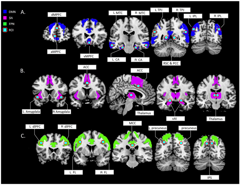
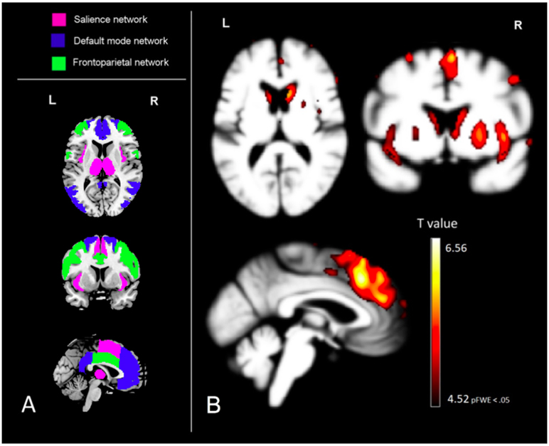
Interpreting others' beliefs, desires and intentions is known as "theory of mind" (ToM), and is often evaluated using simplified measurement tools, which may not correctly reflect the brain circuits that are required for real-life ToM functioning. We aimed to identify the brain structures necessary to correctly infer intentions from realistic scenarios by administering The Awareness of Social Inference Test, Enriched subtest to 47 patients with behavioural variant frontotemporal dementia, 24 patients with progressive supranuclear palsy syndrome, 31 patients with Alzheimer's syndrome, and 77 older healthy controls. Neuroimaging data was analyzed using voxel based morphometry, and participants' understanding of intentions was correlated with voxel-wise and region-of interest data. We found that structural integrity of the cinguloinsular cortex in the salience network (SN) was more pivotal for accurate ToM than previously described, emphasizing the importance of the SN for selectively recognizing and attending to social cues during ToM inferences.

Keywords: Social cognition; neurodegenerative diseases; neuropsychology; salience network; theory of mind; voxel-based morphometry.


Conflict of interest statement

Disclosure of interest

No potential conflict of interest was reported by the authors.

Figures

Figure 1.
Figure 1.
regions of the neuromorphometrics map selected based on region of interests (cyan dots) from existing intrinsically connected network literature (Andrews-Hanna et al., 2010; Beissner et al., 2013; Dosenbach et al., 2007; Pascual et al., 2015; Seeley et al., 2009; Yeo et al., 2011). A. dMPFC = dorsomedial prefrontal cortex (NM = superior frontal gyrus), aMPFC = anteriomedial prefrontal cortex (NM = medial frontal cortex), vMPFC = ventromedial prefrontal cortex (NM = anterior cingulate gyrus), MTC = middle temporal cortex (NM = middle temporal gyrus), CA = hippocampal cortex (NM = parahippocampal gyrus), TPJ = temporoparietal junction (NM = middle occipital gyrus, RSC = retrosplenial cortex (NM = posterior cingulate gyrus), PCC = posterior cingulate cortex (NM = posterior cingulate gyrus), IPL = inforior perietal lobule (NM = angular gyrus). B. Amygdala (NM = amygdala), ACC = anterior cingulate cortex (NM = supplementary motor area), thalamus (NM = thalamus proper), vAI = ventral anterior insula (NM = anterior insula). C. dlPFC = dorsolateral prefrontal cortex (NM = middle frontal gyrus), FL = frontal lobule (NM = precentral gyrus), MCC = middle cingulate cortex (NM = middle cingulate gyrus), precuneus (NM = superior parietal lobule), IPS = inferior periatal sulcus (NM = supramarginal gyrus). DMN = default mode network, SN = salience network, FPN = frontoparietal network, ROI = regions of interest based on functional maxima coordinates , L = left, R = right.
Figure 2.
Figure 2.
Visualization of the voxel-based morphometry derived results of the correlation between gray matter atrophy and the TASIT SI-E ‘do’ score, together with the region of interests for comparison (left). A. region of interests were derived from the Neuromorphometrics atlas. B. Voxel-wise patterns of grey matter volume in which less volume significantly predicts worse performance on the TASIT SI-E ‘do’ score; analyses controlled for age, sex, total intracranial volume, magnet strength, and MMSE score. These results are displayed at a family-wise corrected significance level of pFWE < .05, which corresponds with a minimum T value of 4.52. The same MNI coordinates are used for both image A and B; x= −1, y= 8, z= 10.

References

    1. Abu-Akel A, & Shamay Tsoory S (2011). Neuroanatomical and neurochemical bases of theory of mind. Neuropsychologia, 49(11), 2971–2984. 10.1016/j.neuropsychologia.2011.07.012 - DOI - PubMed
    1. Acosta-Cabronero J, Williams GB, Pereira JMS, Pengas G, & Nestor PJ (2007). The impact of skull-stripping and radio-frequency bias correction on grey-matter segmentation for voxel-based morphometry. NeuroImage, 39(4), 1654–1665. 10.1016/j.neuroimage.2007.10.051 - DOI - PubMed
    1. Amodio DM, & Frith CD (2006). Meeting of minds: the medial frontal cortex and social cognition. Nature Reviews.Neuroscience, 7(4), 268–277. 10.1038/nrn1884 - DOI - PubMed
    1. Andrews-Hanna JR, Reidler JS, Sepulcre J, Poulin R, & Buckner RL (2010). Functional-Anatomic Fractionation of the Brain’s Default Network. Neuron, 65(4), 550–562. 10.1016/j.neuron.2010.02.005 - DOI - PMC - PubMed
    1. Andrews-Hanna JR, Smallwood J, & Spreng RN (2014). The default network and self-generated thought: Component processes, dynamic control, and clinical relevance. Annals of the New York Academy of Sciences, 1316(1), 29–52. 10.1111/nyas.12360 - DOI - PMC - PubMed

Publication types

MeSH terms