Multiple desmoplastic Spitz nevi with BRAF fusions in a patient with ring chromosome 7 syndrome
- PMID: 33522711
- PMCID: PMC9351601
- DOI: 10.1111/pcmr.12963
Multiple desmoplastic Spitz nevi with BRAF fusions in a patient with ring chromosome 7 syndrome
Abstract
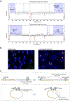
Patients with non-supernumerary ring chromosome 7 syndrome have an increased incidence of hemangiomas, café-au-lait spots, and melanocytic nevi. The mechanism for the increased incidence of these benign neoplasms is unknown. We present the case of a 22-year-old man with ring chromosome 7 and multiple melanocytic nevi. Two nevi, one on the right ear and the other on the right knee, were biopsied and diagnosed as desmoplastic Spitz nevi. Upon targeted next-generation DNA sequencing, both harbored BRAF fusions. Copy number alterations and fluorescence in situ hybridization (FISH) for BRAF suggested that the fusions arose on the ring chromosome 7. Hence, one reason for increased numbers of nevi in patients with non-supernumerary ring chromosome 7 syndrome may be increased likelihood of BRAF fusions, due to the instability of the ring chromosome.
Keywords: BRAF fusion; BRAF gene; Spitz nevus; Spitz tumor; melanocytic nevi; melanocytic nevus; ring chromosome 7; ring chromosome seven; spitzoid.
© 2021 John Wiley & Sons A/S. Published by John Wiley & Sons Ltd.
Conflict of interest statement
Figures


Similar articles
-
BRAF and NRAS mutations in spitzoid melanocytic lesions.Mod Pathol. 2006 Oct;19(10):1324-32. doi: 10.1038/modpathol.3800653. Epub 2006 Jun 23. Mod Pathol. 2006. PMID: 16799476
-
BRAF fusion Spitz neoplasms; clinical morphological, and genomic findings in six cases.J Cutan Pathol. 2020 Dec;47(12):1132-1142. doi: 10.1111/cup.13842. Epub 2020 Sep 14. J Cutan Pathol. 2020. PMID: 32776349
-
Spitz melanoma is a distinct subset of spitzoid melanoma.Mod Pathol. 2020 Jun;33(6):1122-1134. doi: 10.1038/s41379-019-0445-z. Epub 2020 Jan 3. Mod Pathol. 2020. PMID: 31900433 Free PMC article.
-
The role of gene fusions in melanocytic neoplasms.J Cutan Pathol. 2019 Nov;46(11):878-887. doi: 10.1111/cup.13521. Epub 2019 Jun 20. J Cutan Pathol. 2019. PMID: 31152596 Review.
-
[Morphological and genetic aspects of Spitz tumors].Pathologe. 2015 Feb;36(1):37-43, 45. doi: 10.1007/s00292-014-1984-1. Pathologe. 2015. PMID: 25613920 Review. German.
Cited by
-
Top 10 Differential Diagnoses for Desmoplastic Melanoma.Head Neck Pathol. 2023 Mar;17(1):143-153. doi: 10.1007/s12105-023-01536-y. Epub 2023 Mar 16. Head Neck Pathol. 2023. PMID: 36928737 Free PMC article. Review.
-
Kinase Fusions in Spitz Melanocytic Tumors: The Past, the Present, and the Future.Dermatopathology (Basel). 2024 Feb 14;11(1):112-123. doi: 10.3390/dermatopathology11010010. Dermatopathology (Basel). 2024. PMID: 38390852 Free PMC article. Review.
-
Congenital melanocytic naevi initiated by BRAF fusion oncogene with firmness, pruritus and desmoplastic stroma.Br J Dermatol. 2025 Jul 17;193(2):232-239. doi: 10.1093/bjd/ljaf061. Br J Dermatol. 2025. PMID: 40036344 Free PMC article.
References
-
- Botton T, Talevich E, Mishra VK, Zhang T, Shain AH, Berquet C, Gagnon A, Judson RL, Ballotti R, Ribas A, Herlyn M, Rocchi S, Brown KM, Hayward NK, Yeh I, & Bastian BC (2019). Genetic Heterogeneity of BRAF Fusion Kinases in Melanoma Affects Drug Responses. Cell Reports, 29(3), 573–588.e7. 10.1016/j.celrep.2019.09.009 - DOI - PMC - PubMed
-
- Botton T, Yeh I, Nelson T, Vemula SS, Sparatta A, Garrido MC, Allegra M, Rocchi S, Bahadoran P, McCalmont TH, LeBoit PE, Burton EA, Bollag G, Ballotti R, & Bastian BC (2013). Recurrent BRAF kinase fusions in melanocytic tumors offer an opportunity for targeted therapy. Pigment Cell & Melanoma Research, 26(6), 845–851. 10.1111/pcmr.12148 - DOI - PMC - PubMed
-
- Elder DE, Massi D, Scolyer R, & Willemze R (2018). WHO Classification of Skin Tumours (4th Edition). IARC Press.
Publication types
MeSH terms
Substances
Supplementary concepts
Grants and funding
LinkOut - more resources
Full Text Sources
Other Literature Sources
Medical
Research Materials

