. 2021 Feb 17;12(4):782-798.
doi: 10.1021/acschemneuro.1c00007. Epub 2021 Feb 1.

Mass Spectrometry Quantification, Localization, and Discovery of Feeding-Related Neuropeptides in Cancer borealis

Affiliations

Mass Spectrometry Quantification, Localization, and Discovery of Feeding-Related Neuropeptides in Cancer borealis

Kellen DeLaney et al. ACS Chem Neurosci. .

Abstract

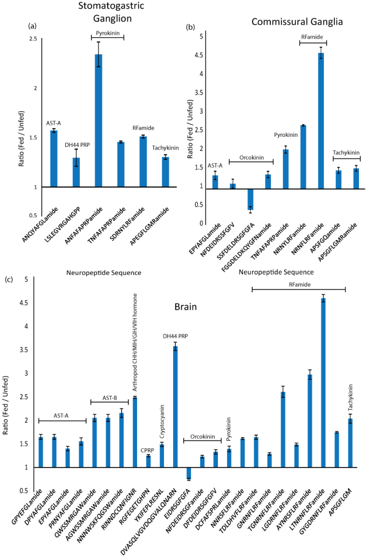
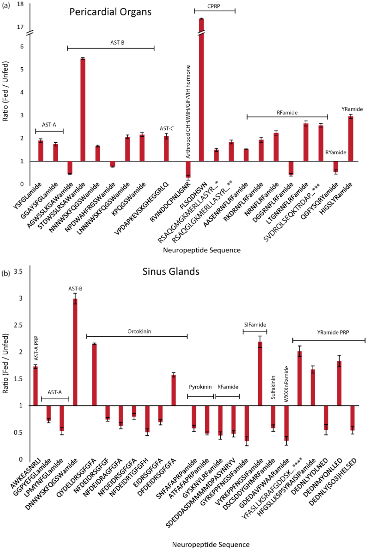
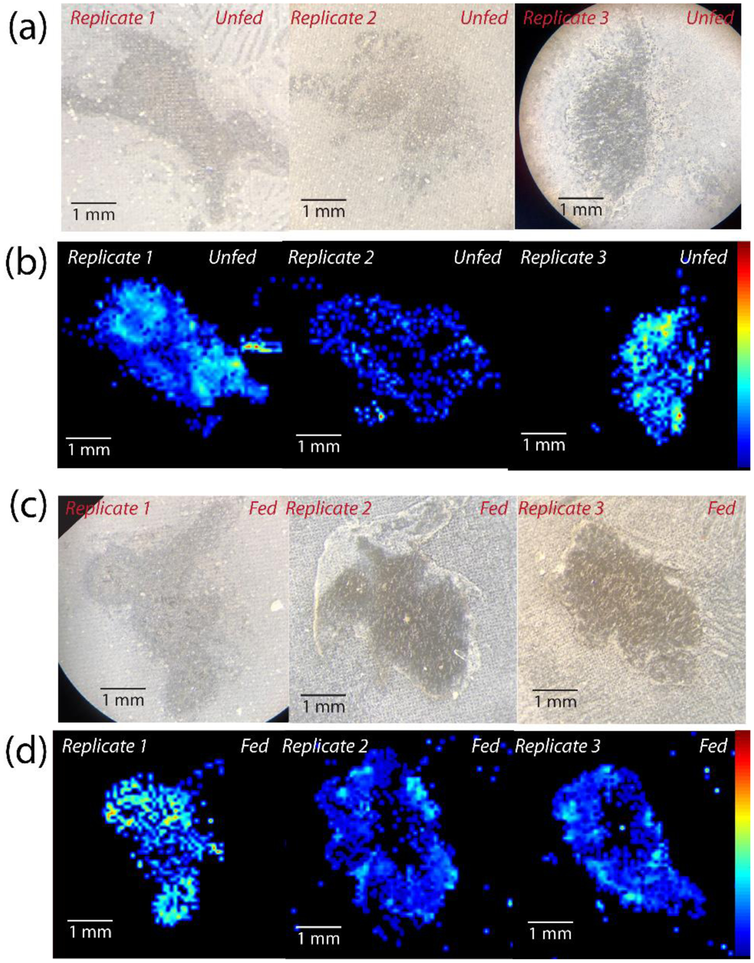
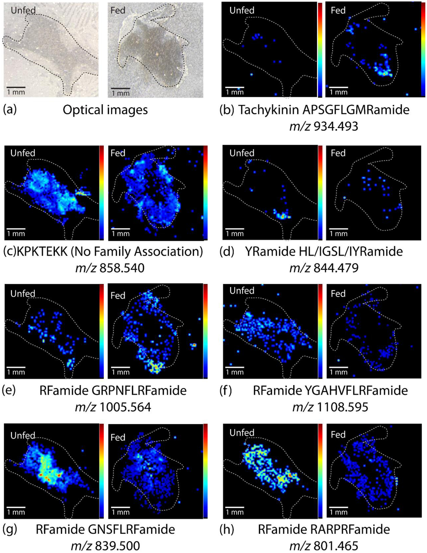
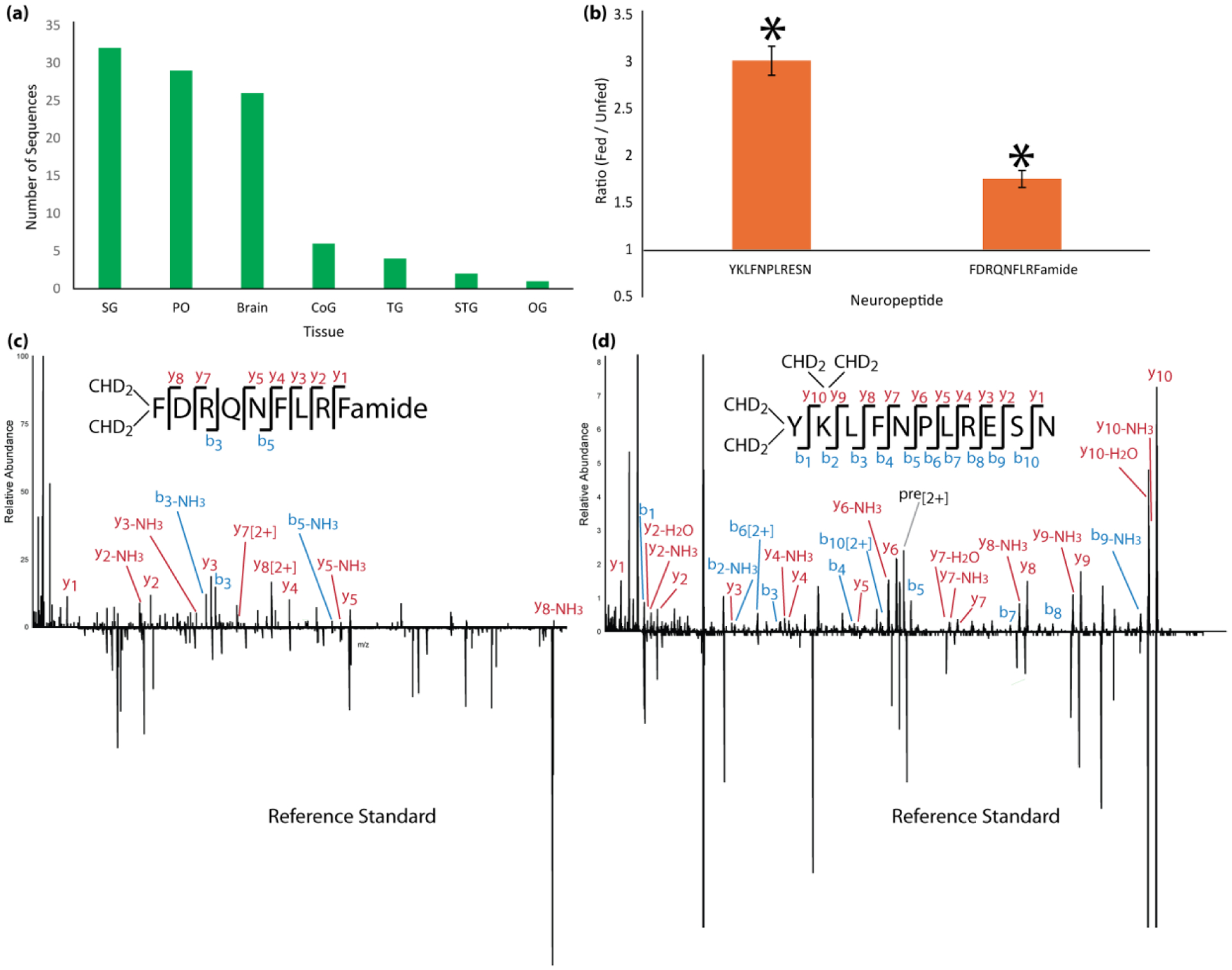
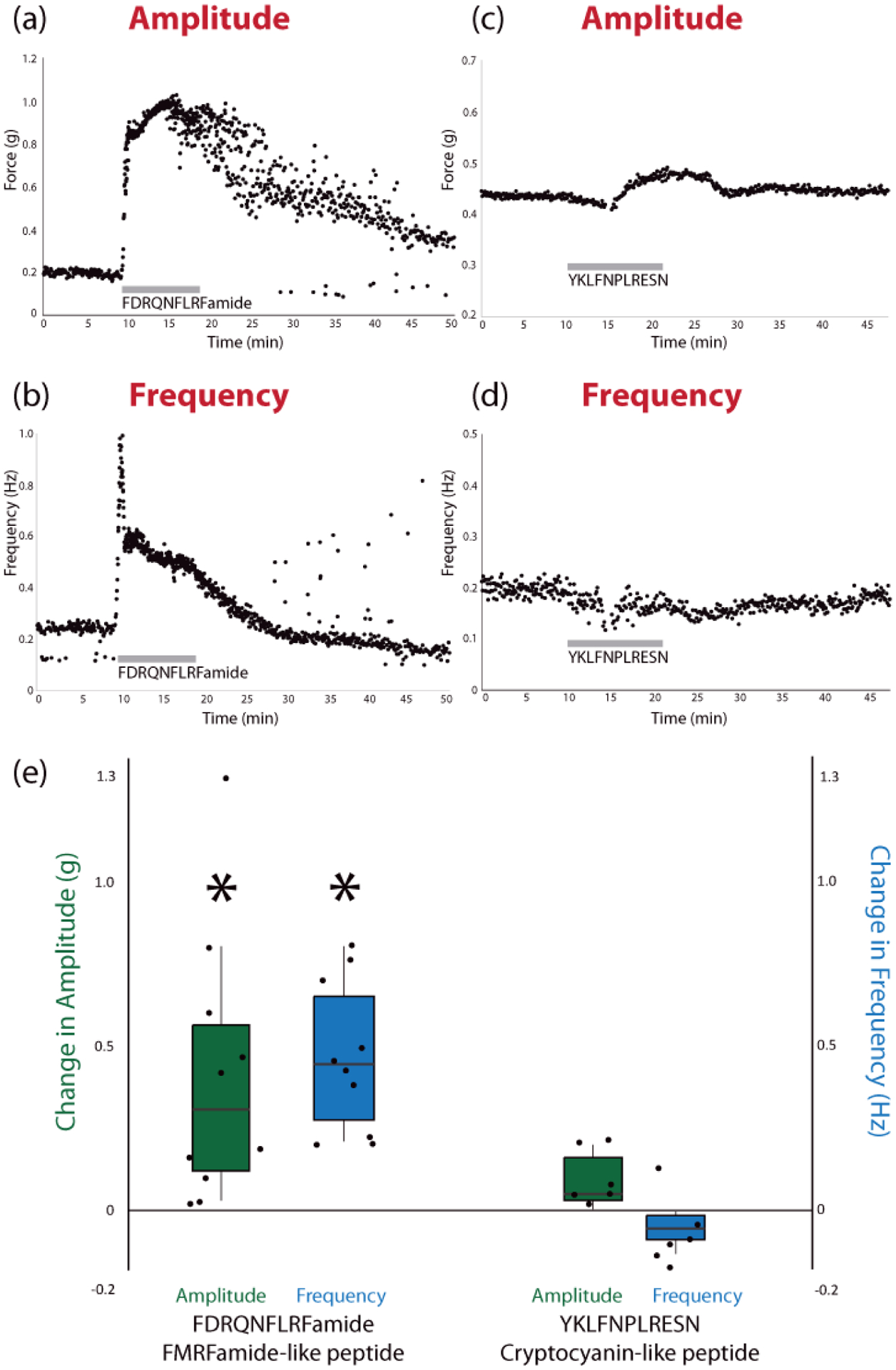
The crab Cancer borealis nervous system is an important model for understanding neural circuit dynamics and modulation, but the identity of neuromodulatory substances and their influence on circuit dynamics in this system remains incomplete, particularly with respect to behavioral state-dependent modulation. Therefore, we used a multifaceted mass spectrometry (MS) method to identify neuropeptides that differentiate the unfed and fed states. Duplex stable isotope labeling revealed that the abundance of 80 of 278 identified neuropeptides was distinct in ganglia and/or neurohemal tissue from fed vs unfed animals. MS imaging revealed that an additional 7 and 11 neuropeptides exhibited altered spatial distributions in the brain and the neuroendocrine pericardial organs (POs), respectively, during these two feeding states. Furthermore, de novo sequencing yielded 69 newly identified putative neuropeptides that may influence feeding state-related neuromodulation. Two of these latter neuropeptides were determined to be upregulated in PO tissue from fed crabs, and one of these two peptides influenced heartbeat in ex vivo preparations. Overall, the results presented here identify a cohort of neuropeptides that are poised to influence feeding-related behaviors, providing valuable opportunities for future functional studies.

Keywords: Mass spectrometry; cardiac neuromodulation; imaging; neuropeptides; stable isotope labeling; stomatogastric nervous system.


Conflict of interest statement

Competing Interests

The authors declare no competing interests.

Figures

Figure 1.
Figure 1.
Neuropeptides exhibiting changes in abundance after feeding, in the stomatogastric ganglion, commissural ganglion and brain of C. borealis. Bar graphs show neuropeptides displaying significantly increased or decreased abundance in 30 minutes post-feeding tissues compared to unfed tissue. The y-axis represents the ratio of fed- to unfed abundance. Significance based on p < 0.05, Unpaired, Two-Tailed Student’s t-Test, n=5. Error bars represent SEM. Neuropeptide families are indicated, allatostatin (AST), crustacean hyperglycemic hormone precursor related peptide (CPRP), precursor related peptide (PRP), diuretic hormone (DH).
Figure 2.
Figure 2.
Feeding state-related changes in neuropeptide abundance in the POs and SGs, two regions of the C. borealis neuroendocrine system. Bar graphs show neuropeptides that were significantly increased or decreased in fed- compared to unfed tissue. The y-axis represents the ratio of fed- to unfed abundance. Significance based on p < 0.05, Unpaired Two-Tailed Student’s t-Test, n=5. Error bars represent SEM. Neuropeptide families are indicated, allatostatin (AST), crustacean hyperglycemic hormone precursor related peptide (CPRP), precursor related peptide (PRP), diuretic hormone (DH), pigment dispersing hormone (PDH). Full sequences: *RSAQGLGKMERLLASYRGALEPNTPLGDLSGSLGHPVE, **RSAQGLGKMEHLLASYRGALEPNTPLGDLSGSLGHPVE, ***SVDRQLSEQKTRDAPVAPTATIHSPAKTQESHRS, ****YFASLLKSRAFGDDSKLIPHNAAGDSEPHLQ
Figure 3.
Figure 3.
Optical and MALDI-MS images of fed and unfed brain sections for all 3 biological replicates of each condition for one representative peptide, KPKTEKK (m/z 858.540), showing the biological reproducibility between sections originating from distinct animals, including (a) optical images of 3 unfed brain sections, (b) MALDI-MS images of unfed brain sections, (c) optical images of fed brain sections, and (d) MALDI-MS images of fed brain sections. Brain sections are oriented such that the anterior is on the right and posterior is on the left.
Figure 4.
Figure 4.
Representative MALDI-MS images of unfed and fed brain sections, including (a) the optical images of the brain section and (b-h) heatmap images of intensity distribution of specific neuropeptides (NPs) within each tissue. All images are normalized to the total ion current (TIC) and oriented such that the anterior side is top right and posterior side is bottom left. The images show differences in neuropeptide distribution between unfed and fed brain tissue for most neuropeptides. White dotted lines indicate approximate outline of tissue. Images from all replicates are shown in Fig S1.
Figure 5.
Figure 5.
Representative MALDI-MS images of unfed and fed pericardial organs, including (a) optical images of the PO and (b-l) heatmap images of intensity distribution of specific neuropeptides within each tissue. PO images were selected from 1 of 3 replicates for each m/z. All images are normalized to the total ion current (TIC) and oriented such that the anterior side is on top and posterior side is on the bottom. The images show differences between unfed and fed tissue for various neuropeptides, such as distinct differences in presence within the tissue or relative abundance in various areas of the tissue. White dotted lines indicate approximate outline of tissue. Images from all replicates are shown in Fig S1.
Figure 6.
Figure 6.
Summary of results of putative novel neuropeptide sequences identified, including (a) the number of novel neuropeptides identified in each tissue and (b) the relative ratio of two of these neuropeptides in fed and unfed POs that were significantly different (p < 0.05, n=5). Error bars represent standard error of the mean. No other novel sequences showed changes that were statistically significant. (c, d) MS/MS spectra mirror plots of the two novel neuropeptides identified with de novo sequencing, including (c) RFamide peptide FDRQNFLRFamide and (d) Cryptocyanin-like peptide YKLFNPLRESN. The top spectrum shows the experimentally-obtained spectrum of each from tissue extract and the bottom mirrored spectrum shows the spectrum obtained from a commercially-synthesized standard. Most key fragment ions are present in both spectra. The difference in intensity of some of the ions is likely due to ion suppression from interfering artifacts in the sample.
Figure 7.
Figure 7.
Results from functional assessment of synthesized peptides on ex vivo whole heart preparations, including examples of the change in amplitude and frequency during perfusion of each peptide (a-d) and the average change in amplitude and frequency for each peptide (e), relative to controls, across all biological replicates (*p < 0.05, Student’s Paired Two-Tailed t-Test, n = 10 for FDRQNFLRFamide, n = 6 for YKLFNPLRESN). Individual values for control and experimental conditions are shown in Fig S72. The average control amplitude was 0.17 g and the average control frequency was 0.20 Hz across all replicates (n = 15). In the boxplots in (e), the center line indicates the median value, the top and bottom of the box represent the 75% quartile and 25% quartile, respectively, and the upper and lower whiskers indicate the largest and smallest values, respectively, not exceeding 1.5 times the inter-quartile range.

Similar articles

Cited by

References

    1. Zimmerman CA, Knight ZA Layers of signals that regulate appetite. Curr Opin Neurobiol 2020, 64, 79–88. - PMC - PubMed
    1. Wu A, Yu B, Komiyama T Plasticity in olfactory bulb circuits. Curr Opin Neurobiol 2020, 64, 17–23. - PMC - PubMed
    1. Hulse BK, Jayaraman V Mechanisms underlying the neural computation of head direction. Annu Rev Neurosci 2019, 43, 31–54. - PubMed
    1. Currier TA, Nagel KI Multisensory control of navigation in the fruit fly. Curr Opin Neurobiol 2019, 64, 10–16. - PMC - PubMed
    1. Barkan CL, Zornik E Feedback to the future: motor neuron contributions to central pattern generator function. J Exp Biol 2019, 222 (Pt 16), 1–9. - PMC - PubMed

Publication types

LinkOut - more resources