. 2021 Jul 16;42(4):407-435.
doi: 10.1210/endrev/bnab004.

Macrophage Responses to Environmental Stimuli During Homeostasis and Disease

Affiliations

Macrophage Responses to Environmental Stimuli During Homeostasis and Disease

Adil Rasheed et al. Endocr Rev. .

Abstract

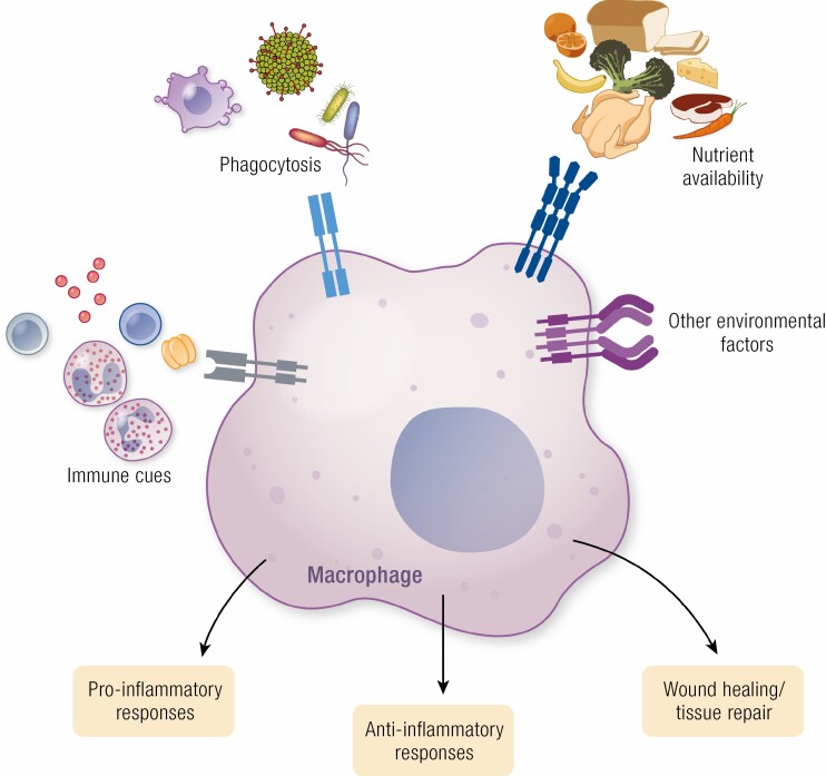
Work over the last 40 years has described macrophages as a heterogeneous population that serve as the frontline surveyors of tissue immunity. As a class, macrophages are found in almost every tissue in the body and as distinct populations within discrete microenvironments in any given tissue. During homeostasis, macrophages protect these tissues by clearing invading foreign bodies and/or mounting immune responses. In addition to varying identities regulated by transcriptional programs shaped by their respective environments, macrophage metabolism serves as an additional regulator to temper responses to extracellular stimuli. The area of research known as "immunometabolism" has been established within the last decade, owing to an increase in studies focusing on the crosstalk between altered metabolism and the regulation of cellular immune processes. From this research, macrophages have emerged as a prime focus of immunometabolic studies, although macrophage metabolism and their immune responses have been studied for centuries. During disease, the metabolic profile of the tissue and/or systemic regulators, such as endocrine factors, become increasingly dysregulated. Owing to these changes, macrophage responses can become skewed to promote further pathophysiologic changes. For instance, during diabetes, obesity, and atherosclerosis, macrophages favor a proinflammatory phenotype; whereas in the tumor microenvironment, macrophages elicit an anti-inflammatory response to enhance tumor growth. Herein we have described how macrophages respond to extracellular cues including inflammatory stimuli, nutrient availability, and endocrine factors that occur during and further promote disease progression.

Keywords: Krebs cycle; epigenetic modifications; immunometabolism; inflammation; macrophage.


Figures

Graphical Abstract
Graphical Abstract
Figure 1.
Figure 1.
Extracellular cues from the environment polarize macrophages. Macrophages express a variety of cell surface receptors that sense extracellular factors that induce macrophage polarization toward a proinflammatory (M1) or anti-inflammatory (M2) state. In cultures, proinflammatory macrophages are polarized by stimulation with lipopolysaccharide (LPS) and interferon γ (IFN-γ). The proinflammatory phenotype of macrophages can also be stimulated by detection of danger-associated molecular patterns (DAMPs)/pattern-associated molecular patterns (PAMPs), such as bacteria, viruses, and exogenous DNA, as well as by cytokines produced by TH1-lymphocytes. Polarization is in part dictated by the activation of transcription factors, such as hypoxia-inducible factor-1α (HIF-1α), IFN-regulatory factor (IRF), and nuclear factor κB (NFκB), and the induction of their proinflammatory gene networks. Furthermore, metabolic reprogramming occurs in these macrophages, primarily by the upregulation of glycolysis. Together these transcriptional and metabolic changes result in inflammasome activation and the production of proinflammatory cytokines and reactive oxygen species. Anti-inflammatory macrophages are produced by the treatment of macrophages with interleukin-4 (IL-4) or IL-13 in vitro and in vivo signals are received from eosinophils and TH2-lymphocytes. Transcription factors required for M2-like polarization include nuclear factor erythroid 2-related factor 2 (NRF2), peroxisome proliferator-activated receptor γ (PPARγ), and signal transducer and activator of transcription 6 (STAT6), leading to the production of anti-inflammatory genes such as IL-10 and arginase 1 (ARG1). In these anti-inflammatory macrophages, metabolism is shifted toward fatty acid oxidation (FAO) and oxidative phosphorylation (OXPHOS). Here, adenosine 5′-monophosphate–activated protein kinase (AMPK) signaling is promoted, and in turn leads to the production of anti-inflammatory cytokines, collagen synthesis, and efferocytosis. Epigenetic modifications also play a role in determining macrophage identity and function, including chromatin remodeling such as acetylation of lysine 27 of histone 3 (H3K27ac) accompanied by binding of transcription factors (LDTF, lineage-determining transcription factors; SDTF, signal-dependent transcription factors), an epigenetic signature that is strongly influenced by environment-derived factors. The plasticity of macrophages between its proinflammatory and anti-inflammatory polarized states can be induced by feedback pathways engaged by the metabolic programs that involve epigenetic changes, such as histone lactylation or demethylation (JMJD3).
Figure 2.
Figure 2.
Macrophage metabolism influences their function. Metabolic changes in macrophages primarily involve alterations to the Krebs cycle. Proinflammatory macrophages (red) and anti-inflammatory macrophages (blue) favor specific pathways that influence their function or inflammatory state. Proinflammatory macrophages favor glycolysis and fatty acid and cholesterol synthesis pathways, whereas anti-inflammatory macrophages prefer fatty acid oxidation. Within the Krebs cycle proinflammatory macrophages exhibit 2 key break points: (1) the inhibition of isocitrate conversion to α-ketoglutarate (by isocitrate dehydrogenase) and the production of itaconate and (2) the inhibition of succinate dehydrogenase, which catalyzes the conversion of succinate to fumarate. This accumulation of succinate allows for the stabilization of the hypoxia-inducible factor-1α (HIF-1α) and the transcription of proinflammatory genes. Succinate metabolism fuels the electron transport chain, which is important for promoting oxidative phosphorylation (OXPHOS) in anti-inflammatory macrophages. However dysfunction in the electron transport chain, including reverse electron transport (RET), produces reactive oxygen species (ROS), characteristic of proinflammatory macrophages. In the urea cycle, arginine metabolism by arginase-1, a prototypical anti-inflammatory marker, produces ornithine, which is a precursor for collagen synthesis used by these macrophages to promote tissue repair and wound healing. Proinflammatory macrophages, on the other hand, express inducible nitric oxide synthase (iNOS), which metabolizes arginine to produce citrulline and nitric oxide (NO), further enhancing the inflammatory status of these macrophages.

References

    1. Davies LC, Jenkins SJ, Allen JE, Taylor PR. Tissue-resident macrophages. Nat Immunol. 2013;14(10):986-995. - PMC - PubMed
    1. Wynn TA, Chawla A, Pollard JW. Macrophage biology in development, homeostasis and disease. Nature. 2013;496(7446): 445-455. - PMC - PubMed
    1. Varol C, Mildner A, Jung S. Macrophages: development and tissue specialization. Annu Rev Immunol. 2015;33:643-675. - PubMed
    1. Janeway CA Jr, Medzhitov R. Innate immune recognition. Annu Rev Immunol. 2002;20:197-216. - PubMed
    1. Taylor PR, Martinez-Pomares L, Stacey M, Lin HH, Brown GD, Gordon S. Macrophage receptors and immune recognition. Annu Rev Immunol. 2005;23:901-944. - PubMed

Publication types

Grants and funding