Review
. 2021 Mar;35(2):125-146.
doi: 10.1007/s40259-020-00465-4. Epub 2021 Feb 1.

Immunogenicity Challenges Associated with Subcutaneous Delivery of Therapeutic Proteins

Affiliations
Review

Immunogenicity Challenges Associated with Subcutaneous Delivery of Therapeutic Proteins

Nicole L Jarvi et al. BioDrugs. 2021 Mar.

Abstract

The subcutaneous route of administration has provided convenient and non-inferior delivery of therapeutic proteins compared to intravenous infusion, but there is potential for enhanced immunogenicity toward subcutaneously administered proteins in a subset of patients. Unwanted anti-drug antibody response toward proteins or monoclonal antibodies upon repeated administration is shown to impact the pharmacokinetics and efficacy of multiple biologics. Unique immunogenicity challenges of the subcutaneous route have been realized through various preclinical and clinical examples, although subcutaneous delivery has often demonstrated comparable immunogenicity to intravenous administration. Beyond route of administration as a treatment-related factor of immunogenicity, certain product-related risk factors are particularly relevant to subcutaneously administered proteins. This review attempts to provide an overview of the mechanism of immune response toward proteins administered subcutaneously (subcutaneous proteins) and comments on product-related risk factors related to protein structure and stability, dosage form, and aggregation. A two-wave mechanism of antigen presentation in the immune response toward subcutaneous proteins is described, and interaction with dynamic antigen-presenting cells possessing high antigen processing efficiency and migratory activity may drive immunogenicity. Mitigation strategies for immunogenicity are discussed, including those in general use clinically and those currently in development. Mechanistic insights along with consideration of risk factors involved inspire theoretical strategies to provide antigen-specific, long-lasting effects for maintaining the safety and efficacy of therapeutic proteins.


Conflict of interest statement

The authors declare that they have no conflicts of interest.

Figures
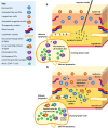
Fig. 1
Fig. 1
Schematic representation of the proposed two-wave mechanism of antigen presentation following subcutaneous injection of protein. a Lymph-borne antigen is delivered to skin-derived LN-resident DCs in DLNs, which begin antigen processing and presentation to antigen-specific naïve CD4+ T cells within T cell areas. b Skin-derived migratory DCs and LCs are recruited into the injection site to acquire antigen. Upregulation of chemokine receptors, CCR7 and CXCR4, on dermal DCs and LCs drives cell migration to initial lymphatics and DLNs. Antigen-loaded migratory DCs and LCs arrive in DLNs for the second wave of presentation to antigen-specific naïve CD4+ T cells, and migratory DCs also transfer antigen to LN-resident DCs [25]. DC dendritic cell, DLN draining lymph node, LC Langerhans cell, LN lymph node
Fig. 2
Fig. 2
Product-related risk factors for immunogenicity of subcutaneously administered therapeutic proteins. Structural or conformational modifications related to instability pathways or proteolytic degradation could generate new/modified epitopes. Protein aggregates or precipitates present in the formulation or formed post-injection can have longer SC retention time. Charge interactions between slight positive charge on mAbs at local physiological pH and negative charge density in ECM may increase SC retention time. Enhanced retention time of protein could confer immunogenic risk by increasing opportunities for encounter with invading dermal DCs and LCs post-injection. Innate immune stimulation by adjuvant-like drug product impurities (e.g., host cell proteins, leachates, and endotoxins) at the injection site can trigger maturation and migration of dermal DCs and LCs. Ag antigen, DC dendritic cell, ECM extracellular matrix, LC Langerhans cell, LN lymph node, mAb monoclonal antibody, SC subcutaneous
Fig. 3
Fig. 3
Selection of existing and theoretical strategies to minimize immunogenicity of subcutaneously administered therapeutic proteins. a Conventional strategies rely on nonspecific immune suppression using small molecule drugs, such as methotrexate, rapamycin, bortezomib, and cyclophosphamide. A combination approach uses the lymphocyte depletion agent rituximab (anti-CD20) with methotrexate and intravenous Ig. b Example lymphocyte modulation strategies are anti-CD3 antibody, engineered antigen-specific FoxP3+ Treg cells, and cytotoxic BAR CD8+ T cells. c Reduction of product-related factors by ex vivo human cell-based assay screening, removal of non-native IgG aggregate precursors by specific absorption to AF.2A1 magnetic beads, and chaperone molecules to improve protein stability. d Inhibition of CCL19/CCL21 directed DC migration to lymph nodes by the small molecule CCR7 inhibitor cosalane (anti-HIV agent). e mAb humanization by incorporation of fully human content apart from complementarity-determining regions. In silico prediction of T or B cell epitopes on proteins to perform de-immunization or incorporation of Treg cell epitopes (Tregitopes). f Peripheral tolerance induction by co-administration of OPLS with therapeutic protein subcutaneously to induce tolerogenic DCs and antigen-specific Treg responses [7, 178, 179, 183, 185, 186, 191, 192, 197, 209]. BAR B cell antibody-targeting receptor, CDR complementarity determining region, DC dendritic cell, DLN draining lymph node, FoxP3 forkhead box P3, HIV human immunodeficiency virus, IDO indoleamine 2,3-dioxygenase, Ig immunoglobulin, IL interleukin, mAb monoclonal antibody, MHC major histocompatibility complex, OPLS O-phospho-L-serine, RA retinoic acid, TCR T cell receptor, TGFβ tumor growth factor-β, Treg regulatory T cell
Fig. 4
Fig. 4
Immune tolerance induction using SC co-administration of OPLS and therapeutic protein to mitigate immunogenicity. Top: (1) Uptake and processing of protein by skin-derived immature DCs licenses DC maturation and migration to DLNs. (2) DCs present peptide:MHC II complexes to naïve CD4+ T cells in T cell areas and induce (3) differentiation and proliferation of effector CD4+ T cells. (4) B cell activation and differentiation in germinal centers generates (5) memory B cells and plasma cells producing ADA (e.g., IgG). Bottom: (1) FVIII and OPLS are mixed immediately before SC administration. Uptake and processing of FVIII in the presence of OPLS by skin-derived immature DCs induces tolerogenic DCs, with downregulation of proinflammatory cytokine production and co-stimulatory molecule expression, but similar MHC II expression compared to mature DCs. (2) Tolerogenic DCs retain migratory activity and reach DLNs to present peptide:MHC II complexes to naïve CD4+ T cells in T cell areas. (3) Antigen presentation in the context of regulatory mediators with low co-stimulatory signals induces Treg cells. (4) Treg cells can suppress effector T cell function via metabolic disruption, adenosine A2A receptor activation, cytolysis, and deprivation of crucial cytokines (e.g. IL-2). (5) Treg cells produce key regulatory cytokines. (6) Interaction of Treg CTLA-4 with DC co-stimulatory molecule B7 induces tolerogenic DCs that suppress effector T cells via regulatory mediators [–204, 209]. Note that steps 4–6 (bottom) represent general pathways of tolerance induction by Treg cells; the mechanism of OPLS-mediated hyporesponsiveness beyond tolerogenic DCs requires further investigation. ADA anti-drug antibody, CD40L CD40 ligand, CTLA-4 cytotoxic T lymphocyte-associated protein 4, DC dendritic cell, DLN draining lymph node, FVIII factor VIII, IDO indoleamine 2,3-dioxygenase, Ig immunoglobulin, IL interleukin, MHC II major histocompatibility complex II, OPLS O-phospho-L-serine, RA retinoic acid, SC subcutaneous, TCR T cell receptor, TGFβ tumor growth factor-β, TNFα tumor necrosis factor-α, Treg regulatory T cell

References

    1. U.S. Food and Drug Administration. Guidance for Industry: Immunogenicity Assessment for Therapeutic Protein Products. Silver Spring, MD: FDA; 2014. https://www.fda.gov/downloads/drugs/guidances/ucm338856.pdf Accessed 12 Jun 2020.
    1. De Groot AS, Scott DW. Immunogenicity of protein therapeutics. Trends Immunol. 2007;28(11):482–490. doi: 10.1016/j.it.2007.07.011. - DOI - PubMed
    1. Lee CA, Kessler CM, Varon D, Martinowitz U, Heim M, Jacquemin MG, et al. Factor VIII immunogenicity. Haemophilia. 1998;4(4):552–557. doi: 10.1046/j.1365-2516.1998.440552.x. - DOI - PubMed
    1. Tieu P, Chan A, Matino D. Molecular mechanisms of inhibitor development in hemophilia. Mediterr J Hematol Infect Dis. 2020;12(1):e2020001. doi: 10.4084/mjhid.2020.001. - DOI - PMC - PubMed
    1. Lee CA, Kessler CM, Varon D, Martinowitz U, Heim M, Hay CRM. Factor VIII inhibitors in mild and moderate-severity haemophilia A. Haemophilia. 1998;4(4):558–563. doi: 10.1046/j.1365-2516.1998.440558.x. - DOI - PubMed