. 2021 Mar;25(6):2944-2955.
doi: 10.1111/jcmm.16329. Epub 2021 Feb 1.

Mangiferin prevents myocardial infarction-induced apoptosis and heart failure in mice by activating the Sirt1/FoxO3a pathway

Affiliations

Mangiferin prevents myocardial infarction-induced apoptosis and heart failure in mice by activating the Sirt1/FoxO3a pathway

Lingli Chen et al. J Cell Mol Med. 2021 Mar.

Abstract

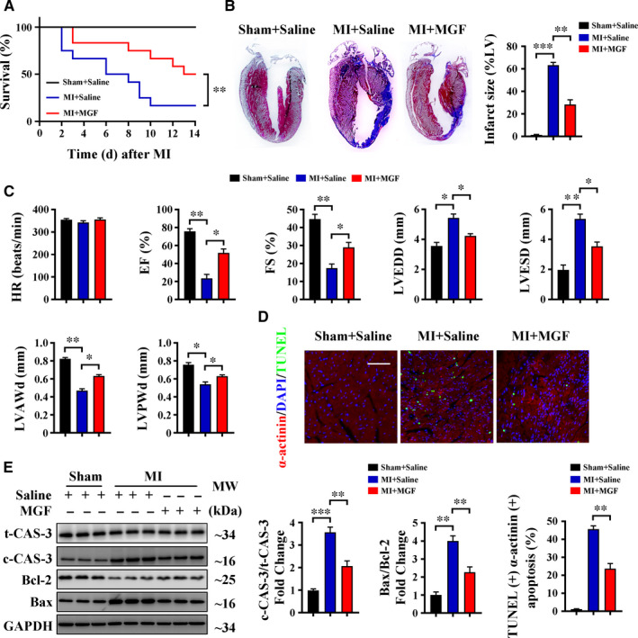
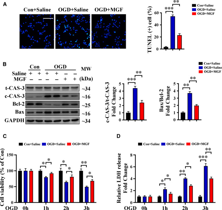
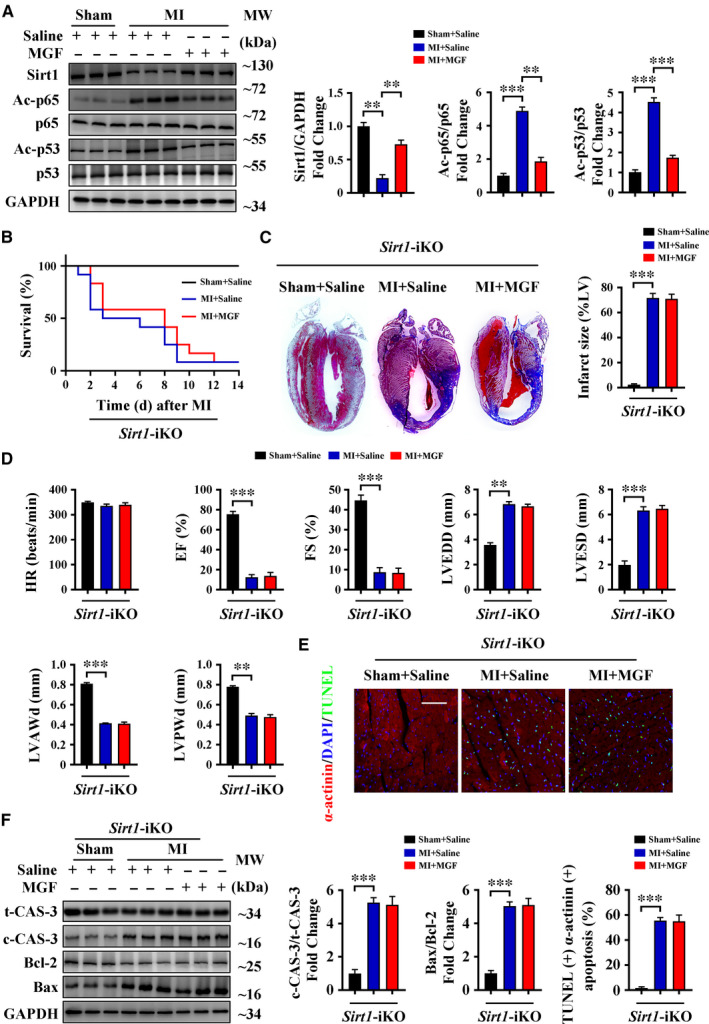
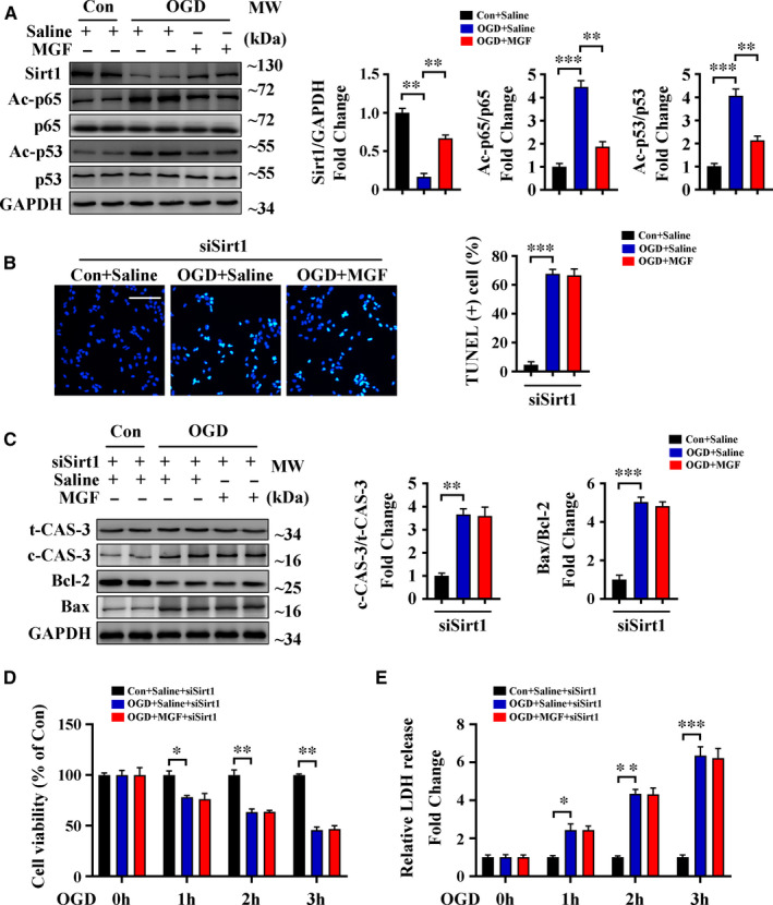
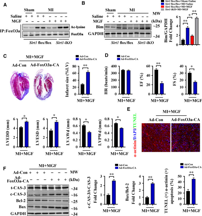
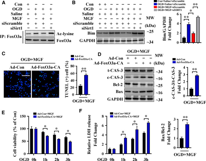
Myocardial infarction (MI) commonly leads to cardiomyocyte apoptosis and heart failure. Mangiferin is a natural glucosylxanthone extracted from mango fruits and leaves, which has anti-apoptotic and anti-inflammatory properties in experimental cardiovascular diseases. In the present study, we investigated the role and detailed mechanism of mangiferin in MI. We used ligation of the left anterior descending coronary artery to establish an MI model in vivo, and cardiomyocyte-specific Sirt1 knockout mice were used to identify the mechanism of mangiferin. For in vitro studies, oxygen and glucose deprivation (OGD) was used to mimic ischaemia in H9c2 cardiomyocytes. In mice, mangiferin treatment increased Sirt1 expression after MI, significantly reduced the infarct area, and prevented MI-induced apoptosis and heart failure. Mangiferin reduced OGD-induced cellular apoptosis in H9c2 cells. Meanwhile, Sirt1 knockout/silencing abolished the protective effects of mangiferin. Further studies revealed that mangiferin increased FoxO3a deacetylation by up-regulating Sirt1, thus preventing apoptosis, and adenovirus-mediated constitutive acetylation of FoxO3a restricted the anti-apoptotic effects of mangiferin in vivo and in vitro. Our results indicate that mangiferin prevents cardiomyocyte apoptosis and the subsequent heart failure by activating the Sirt1/FoxO3a pathway in MI, and suggest that mangiferin may have an interesting potential in following studies towards clinical evaluation.

Keywords: FoxO3a; apoptosis; mangiferin; myocardial infarction; sirt1.


Conflict of interest statement

All authors declare that there are no conflicts of interest in this article.

Figures

FIGURE 1
FIGURE 1
Mangiferin alleviates MI‐induced cardiomyocyte apoptosis and heart failure. (A‐E) C57BL/6 mice were subjected to sham or LAD ligation followed by the treatment with or without mangiferin for 14 d. A, Kaplan‐Meier survival curves of different groups of mice (n = 15 mice in each group). B, Representative images of Masson's trichrome staining of the heart sections and macroscopic measurements of the infarct size. C, M‐mode echocardiography data for HR, EF (%), FS (%), LVEDD, LVESD, LVAWd and LVPWd. D, Representative confocal scans for α‐actinin, TUNEL and DAPI staining (red, green and blue, respectively) of the heart sections and quantitative analysis (lower panel) of TUNEL + and α‐actinin + cells, Scale bar = 100 μm. E, Western blotting assay and quantitative analysis of cleaved caspase 3 expression and Bax/Bcl‐2 ratio in the heart tissue homogenates of different groups of mice. Unless otherwise stated, data are mean ± SEM for n = 6 mice in each group. *P < .05; **P < .01; ***P < .001. c‐CAS‐3, cleaved caspase 3; EF, ejection fraction; FS, fractional shortening; HR, heart rate; LVAWd, left ventricular anterior wall diastolic thickness; LVEDD, left ventricular end‐diastolic diameter; LVESD, left ventricular end‐systolic diameter; LVPWd, left ventricular posterior wall diastolic thickness; MGF, mangiferin; t‐CAS‐3, total caspase 3
FIGURE 2
FIGURE 2
Mangiferin alleviates OGD‐induced cell apoptosis and improves cell viability. A‐D, H9c2 cardiomyocytes were subjected to control operation or OGD for 3 h with or without mangiferin treatment. A, Representative confocal scans for TUNEL and DAPI staining (green and blue, respectively) of the fixed cells and quantitative analysis of TUNEL + cells, Scale bar = 100 μm. B, Western blotting assay and quantitative analysis of cleaved caspase 3 expression and Bax/Bcl‐2 ratio in different groups of cells. C, MTT assay for the detection of cell viability in different groups of cells at indicated time‐points. D, LDH release assessment for the detection of cell injury in different groups of cells at indicated time‐points. Data are mean ± SEM for 3 independent experiments. *P < .05; **P < .01; ***P < .001. Con, control
FIGURE 3
FIGURE 3
Sirt1 is essential for the protective effects of mangiferin during MI. A, C57BL/6 mice were subjected to sham or LAD ligation followed by the treatment with or without mangiferin for 14 d, Western blotting assay and quantitative analysis of Sirt1, acetylated p65 (Ac‐p65), p65, acetylated p53 (Ac‐p53) and p53 in the heart tissue homogenates of different groups of mice. B‐F, Sirt1‐iKO mice were subjected to sham or LAD ligation followed by the treatment with or without mangiferin for 14 d. B, Kaplan‐Meier survival curves of different groups of mice (n = 15 mice in each group). C, Representative images of Masson's trichrome staining of the heart sections and macroscopic measurements of the infarct size. D, M‐mode echocardiography data for HR, EF (%), FS (%), LVEDD, LVESD, LVAWd and LVPWd. E, Representative confocal scans for α‐actinin, TUNEL and DAPI staining (red, green and blue, respectively) of the heart sections and quantitative analysis (lower panel) of TUNEL + and α‐actinin + cells, Scale bar = 100 μm. F, Western blotting assay and quantitative analysis of cleaved caspase 3 expression and Bax/Bcl‐2 ratio in the heart tissue homogenates of different groups of mice. Unless otherwise stated, data are mean ± SEM for n = 6 mice in each group. *P < .05; **P < .01; ***P < .001
FIGURE 4
FIGURE 4
Sirt1 is essential for the protective effects of mangiferin during OGD. A, H9c2 cardiomyocytes were subjected to control operation or OGD for 3 h with or without mangiferin treatment, Western blotting assay and quantitative analysis of Sirt1, acetylated p65 (Ac‐p65), p65, acetylated p53 (Ac‐p53) and p53 in different groups of cells. (B‐E) H9c2 cardiomyocytes treated with Sirt1 siRNA were subjected to control operation or OGD for 3 h with or without mangiferin treatment. B, Representative confocal scans for TUNEL and DAPI staining (green and blue, respectively) of the fixed cells and quantitative analysis of TUNEL + cells, Scale bar = 100 μm. C, Western blotting assay and quantitative analysis of cleaved caspase 3 expression and Bax/Bcl‐2 ratio in different groups of cells. D, MTT assay for the detection of cell viability in different groups of cells at indicated time‐points. E, LDH release assessment for the detection of cell injury in different groups of cells at indicated time‐points. Data are mean ± SEM for 3 independent experiments. *P < .05; **P < .01; ***P < .001
FIGURE 5
FIGURE 5
Mangiferin protects the heart through Sirt1‐mediated deacetylation of FoxO3a during MI. A‐B, Sirt1 flox/flox and Sirt1‐iKO mice were subjected to sham or LAD ligation followed by the treatment with or without mangiferin for 14 d. A, IP assay showed the acetylation level of FoxO3a in the heart tissue homogenates of different groups of mice. B, Western blotting assay and quantitative analysis of Bim expression in the heart tissue homogenates of different groups of mice. (C‐F) C57BL/6 mice transfected with Ad‐Con or Ad‐FoxO3a‐CA vectors were subjected to LAD ligation followed by the treatment with mangiferin for 14 d. C, Representative images of Masson's trichrome staining of the heart sections and macroscopic measurements of the infarct size. D, M‐mode echocardiography data for HR, EF (%), FS (%), LVEDD, LVESD, LVAWd and LVPWd. E, Representative confocal scans for α‐actinin, TUNEL and DAPI staining (red, green and blue, respectively) of the heart sections and quantitative analysis (lower panel) of TUNEL + and α‐actinin + cells, Scale bar = 100 μm. F, Western blotting assay and quantitative analysis of cleaved caspase 3 expression and Bax/Bcl‐2 ratio in the heart tissue homogenates of different groups of mice. Data are mean ± SEM for n = 6 mice in each group. *P < .05; **P < .01; ***P < .001
FIGURE 6
FIGURE 6
Mangiferin alleviates OGD‐induced cell apoptosis and injury through Sirt1‐mediated deacetylation of FoxO3a. A‐B, H9c2 cardiomyocytes treated with scramble siRNA or Sirt1 siRNA were subjected to control operation or OGD for 3 h with or without mangiferin treatment. A, IP assay showed the acetylation level of FoxO3a in different groups of cells. B, Western blotting assay and quantitative analysis of Bim expression in different groups of cells. C‐F, H9c2 cardiomyocytes transfected with Ad‐Con or Ad‐FoxO3a‐CA vectors were subjected to OGD for 3 h with mangiferin treatment. C, Representative confocal scans for TUNEL and DAPI staining (green and blue, respectively) of the fixed cells and quantitative analysis of TUNEL + cells, Scale bar = 100 μm. D, Western blotting assay and quantitative analysis (right and lower panel) of cleaved caspase 3 expression and Bax/Bcl‐2 ratio in different groups of cells. E, MTT assay for the detection of cell viability in different groups of cells at indicated time‐points. F, LDH release assessment for the detection of cell injury in different groups of cells at indicated time‐points. Data are mean ± SEM for 3 independent experiments. *P < .05; **P < .01; ***P < .001

References

    1. Musher DM, Abers MS, Corrales‐Medina VF. Acute Infection and Myocardial Infarction. N Engl J Med. 2019;380:171‐176. - PubMed
    1. Reed GW, Rossi JE, Cannon CP. Acute myocardial infarction. Lancet. 2017;389:197‐210. - PubMed
    1. Jiang H, Wang H, Liu T, et al. Co‐cultured the MSCs and cardiomyocytes can promote the growth of cardiomyocytes. Cytotechnology. 2018;70:793‐806. - PMC - PubMed
    1. Tzahor E, Poss KD. Cardiac regeneration strategies: Staying young at heart. Science. 2017;356:1035‐1039. - PMC - PubMed
    1. Singh R, Letai A, Sarosiek K. Regulation of apoptosis in health and disease: the balancing act of BCL‐2 family proteins. Nat Rev Mol Cell Biol. 2019;20:175‐193. - PMC - PubMed

Publication types

MeSH terms