Review
. 2021 Feb;9(3):e14726.
doi: 10.14814/phy2.14726.

SARS CoV-2 related microvascular damage and symptoms during and after COVID-19: Consequences of capillary transit-time changes, tissue hypoxia and inflammation

Affiliations
Review

SARS CoV-2 related microvascular damage and symptoms during and after COVID-19: Consequences of capillary transit-time changes, tissue hypoxia and inflammation

Leif Østergaard. Physiol Rep. 2021 Feb.

Abstract

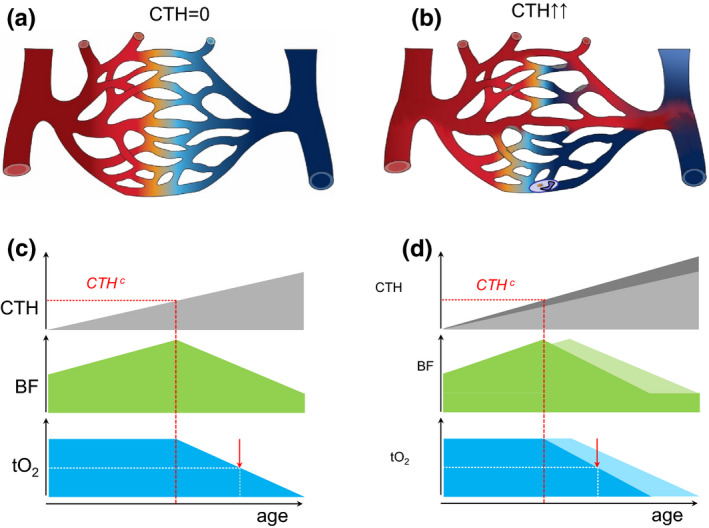
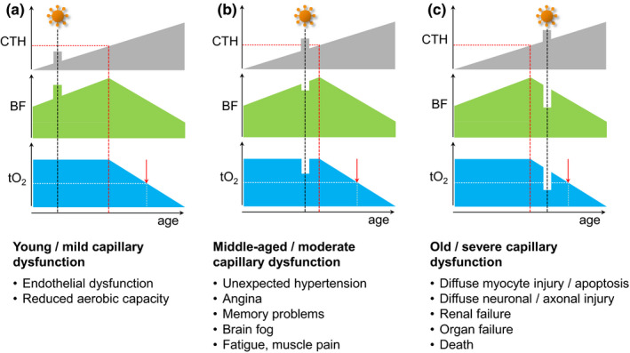
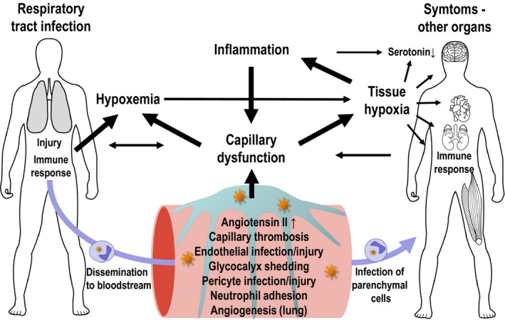
Corona virus disease 2019 (COVID-19) causes symptoms from multiple organs after infection by severe acute respiratory syndrome corona virus 2 (SARS CoV-2). They range from early, low blood oxygen levels (hypoxemia) without breathlessness ("silent hypoxia"), delirium, rashes, and loss of smell (anosmia), to persisting chest pain, muscle weakness and -pain, fatigue, confusion, memory problems and difficulty to concentrate ("brain fog"), mood changes, and unexpected onset of hypertension or diabetes. SARS CoV-2 affects the microcirculation, causing endothelial cell swelling and damage (endotheliitis), microscopic blood clots (microthrombosis), capillary congestion, and damage to pericytes that are integral to capillary integrity and barrier function, tissue repair (angiogenesis), and scar formation. Similar to other instances of critical illness, COVID-19 is also associated with elevated cytokine levels in the systemic circulation. This review examines how capillary damage and inflammation may contribute to these acute and persisting COVID-19 symptoms by interfering with blood and tissue oxygenation and with brain function. Undetectable by current diagnostic methods, capillary flow disturbances limit oxygen diffusion exchange in lungs and tissue and may therefore cause hypoxemia and tissue hypoxia. The review analyzes the combined effects of COVID-19-related capillary damage, pre-existing microvascular changes, and upstream vascular tone on tissue oxygenation in key organs. It identifies a vicious cycle, as infection- and hypoxia-related inflammation cause capillary function to deteriorate, which in turn accelerates hypoxia-related inflammation and tissue damage. Finally, the review addresses the effects of low oxygen and high cytokine levels in brain tissue on neurotransmitter synthesis and mood. Methods to assess capillary functions in human organs and therapeutic means to protect capillary functions and stimulate capillary bed repair may prove important for the individualized management of COVID-19 patients and targeted rehabilitation strategies.

Keywords: COVID-19; brain; capillary dysfunction; heart; hypoxemia; hypoxia; inflammation; long-term symptoms; lungs; microcirculation; muscle.


Conflict of interest statement

LØ is a minority shareholder and Scientific Advisory Board member in Cercare Medical Aps, Denmark and received honoraria for lecturing from Takeda Pharmaceutical Company Limited.

Figures

FIGURE 1
FIGURE 1
Capillary dysfunction and tissue oxygenation (a) Schematic capillary bed. Fully oxygenated blood is indicated by red and its transition to blue illustrates gradual deoxygenation as oxygen diffuses into tissue. In this illustration, the distribution of blood across parallel capillary paths is homogenous (no capillary transit‐time heterogeneity, i.e., CTH=0), providing optimal oxygen extraction. Modified from (Østergaard, 2020). (b) Capillary flow disturbances can be caused by either a narrowing or a widening of individual capillary segments, or by altered blood properties, such as reduced erythrocyte deformability or neutrophil adhesion after glycocalyx shedding. Note how the inability to homogenize capillary transit times across the capillary bed (CTH > 0) leads to poorer oxygen extraction although blood flow is identical to panel a. Modified from (Østergaard, 2020). (c) This panel illustrates how, for modest increases in CTH, tissue oxygen tension (tO2) can be maintained by a compensatory increase in blood flow, contrary to our current vascular disease paradigm (Østergaard, 2020). The dashed red line indicates CTHc, above which metabolic demands can only be met by limiting blood flow (Angleys et al., 2015; Jespersen & Østergaard, 2012). The lower blood supply causes tO2 to decrease, whereas the longer blood transit times and higher blood‐tissue oxygen concentration gradients provide more efficient oxygen extraction from blood, understood as a higher oxygen extraction fraction (OEF). The futility of increasing blood flow beyond the capillary bed's capacity to extract blood's oxygen may be reflected in reperfusion injury after ischemic episodes, during which capillaries irreversibly constrict (O'Farrell et al., 2017; Yemisci et al., 2009). As CTH increases beyond CTHc, BF, and tO2 gradually decreases. The red arrow indicates a 50% reduction in tissue oxygen tension to highlight the threat to critical, ATP‐sensitive cell processes and organ functions. (d) The curves illustrate how cardiovascular risk factors that accelerate capillary injury (Østergaard et al., 2016) modify the curves displayed in panel c (indicated in lighter, gray, green and blue, respectively). Note how CTHc and the onset of critically low tissue oxygen levels are expected to be reached earlier in the affected tissues. The clinical correlates of this earlier attenuation of blood flow may be earlier onset of endothelial dysfunction and hypertension, higher morbidity due to accelerated microvascular injury in critical organs, and lower life expectancy
FIGURE 2
FIGURE 2
Temporary capillary flow disturbances and tissue oxygenation. This figure illustrates how temporary COVID‐19‐related flow disturbances are expected to affect flood flow (BF) and tissue oxygen tension (tO2). Panels show expected responses in tissue with mild (panel a), moderate (panel b), and severe (panel c) pre‐existing capillary dysfunction. (a) If tissue CTH remains below CTHc during COVID‐19‐related capillary flow disturbances, tO2, and thereby organ functions during rest, are likely to remain unaltered. For organs that rely on capillary transit time homogenization to meet their metabolic demands during work, such as the heart and skeletal muscle (Angleys & Østergaard, 2020), the additional capillary dysfunction is expected to reduce maximum aerobic capacity, affecting the performance of, for example, young athletes. These signs are expected to reverse upon successful therapeutic or intrinsic capillary recanalization/repair. Irreversible changes to the capillary bed's oxygen extraction capacity, however, may accelerate the emergence of symptoms and disease changes in the affected tissues, cf. Figure 1d. (b) For patients with asymptomatic, moderate preexisting capillary flow disturbances, a COVID‐19‐related CTH increase may cause them to exceed CTHc and lead to a reduction in blood flow and tissue oxygen tension. While, for example, MCI‐like symptoms are expected to disappear with disease‐related capillary flow disturbances, they may herald developing capillary dysfunction and the benefits of managing cardiovascular risk factors to prevent or delay their recurrence in later life (Hachinski et al., 2019). In COVID‐19 patients with pre‐existing myocardial capillary dysfunction, a sudden CTH increases may reduce tO2 to levels so that they experience angina during exercise in the absence of flow‐limiting coronary disease (Østergaard et al., 2014). (c) In patients with symptomatic, pre‐existing severe capillary dysfunction, further increases in CTH are expected to cause immediate drops in oxygen availability and worsen preexisting symptoms (e.g., confusion or delirium in patients who already have dementia, myocardial damage, or kidney failure). If oxygen levels fall below the metabolic requirement of critical cell functions, tissue and organ damage is expected to cause lasting symptoms or even organ failure and premature death
FIGURE 3
FIGURE 3
Interactions between capillary function, inflammation, hypoxia, and neurotransmission. The expression of ACE2 and other SARS‐CoV‐2 entry factors on parenchymal cells and observations of infected cells in biopsy material hold important clues to understand COVID‐19‐related organ damage

References

    1. Ackermann, M. , Verleden, S. E. , Kuehnel, M. , Haverich, A. , Welte, T. , Laenger, F. , Vanstapel, A. , Werlein, C. , Stark, H. , Tzankov, A. , Li, W. W. , Li, V. W. , Mentzer, S. J. , & Jonigk, D. (2020). Pulmonary vascular endothelialitis, thrombosis, and angiogenesis in Covid‐19. New England Journal of Medicine, 383(2), 120–128. 10.1056/NEJMoa2015432 - DOI - PMC - PubMed
    1. Angleys, H. , & Østergaard, L. (2020). Krogh’s capillary recruitment hypothesis, 100 years on: Is the opening of previously closed capillaries necessary to ensure muscle oxygenation during exercise? American Journal of Physiology‐Heart and Circulatory Physiology, 318(2), H425–H447. 10.1152/ajpheart.00384.2019 - DOI - PubMed
    1. Angleys, H. , Østergaard, L. , & Jespersen, S. N. (2015). The effects of capillary transit time heterogeneity (CTH) on brain oxygenation. Journal of Cerebral Blood Flow & Metabolism, 35(5), 806–817. 10.1038/jcbfm.2014.254 - DOI - PMC - PubMed
    1. Armulik, A. , Genove, G. , & Betsholtz, C. (2011). Pericytes: Developmental, physiological, and pathological perspectives, problems, and promises. Developmental Cell, 21(2), 193–215. 10.1016/j.devcel.2011.07.001 - DOI - PubMed
    1. Armulik, A. , Genove, G. , Mae, M. , Nisancioglu, M. H. , Wallgard, E. , Niaudet, C. , He, L. , Norlin, J. , Lindblom, P. , Strittmatter, K. , Johansson, B. R. , & Betsholtz, C. (2010). Pericytes regulate the blood‐brain barrier. Nature, 468(7323), 557–561. 10.1016/j.devcel.2011.07.001 - DOI - PubMed

Publication types

LinkOut - more resources