. 2021 Jun 15;203(12):1533-1545.
doi: 10.1164/rccm.202003-0719OC.

NEDD9 Is a Novel and Modifiable Mediator of Platelet-Endothelial Adhesion in the Pulmonary Circulation

Affiliations

NEDD9 Is a Novel and Modifiable Mediator of Platelet-Endothelial Adhesion in the Pulmonary Circulation

George A Alba et al. Am J Respir Crit Care Med. .

Abstract

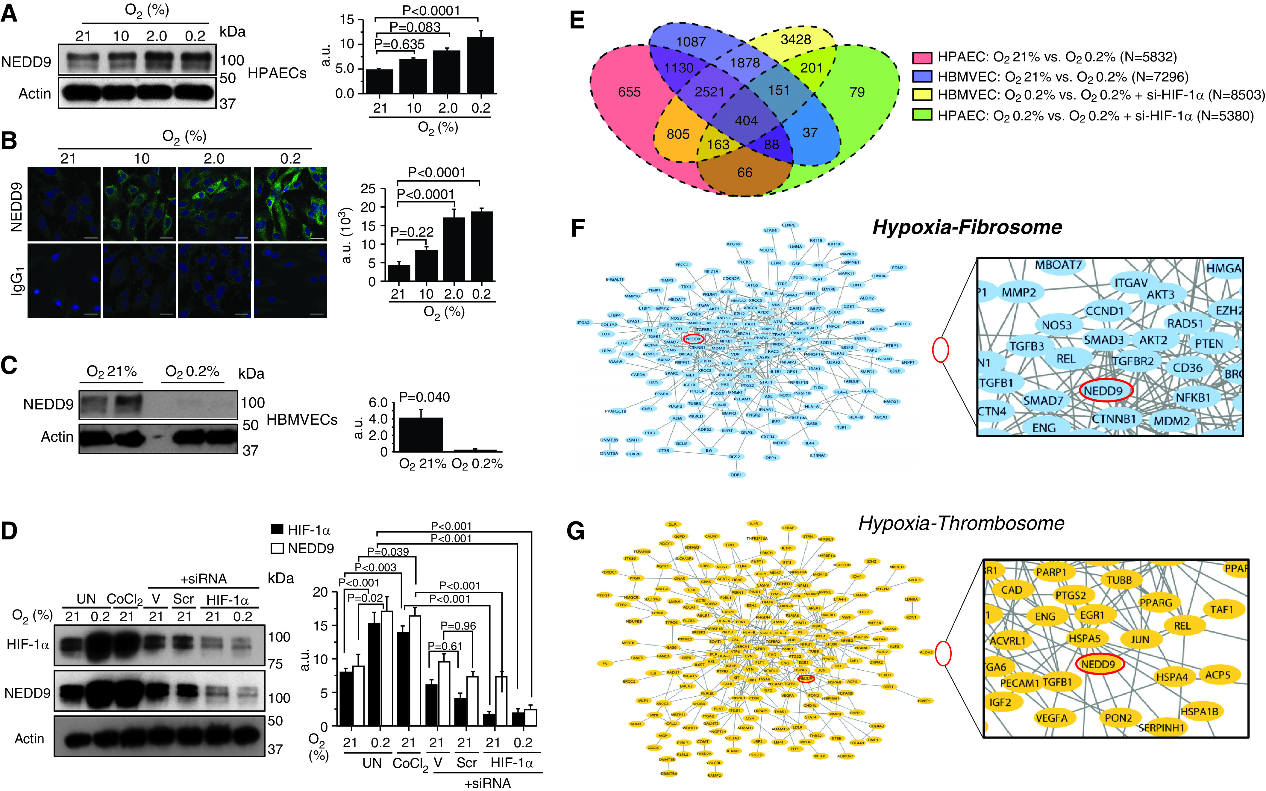
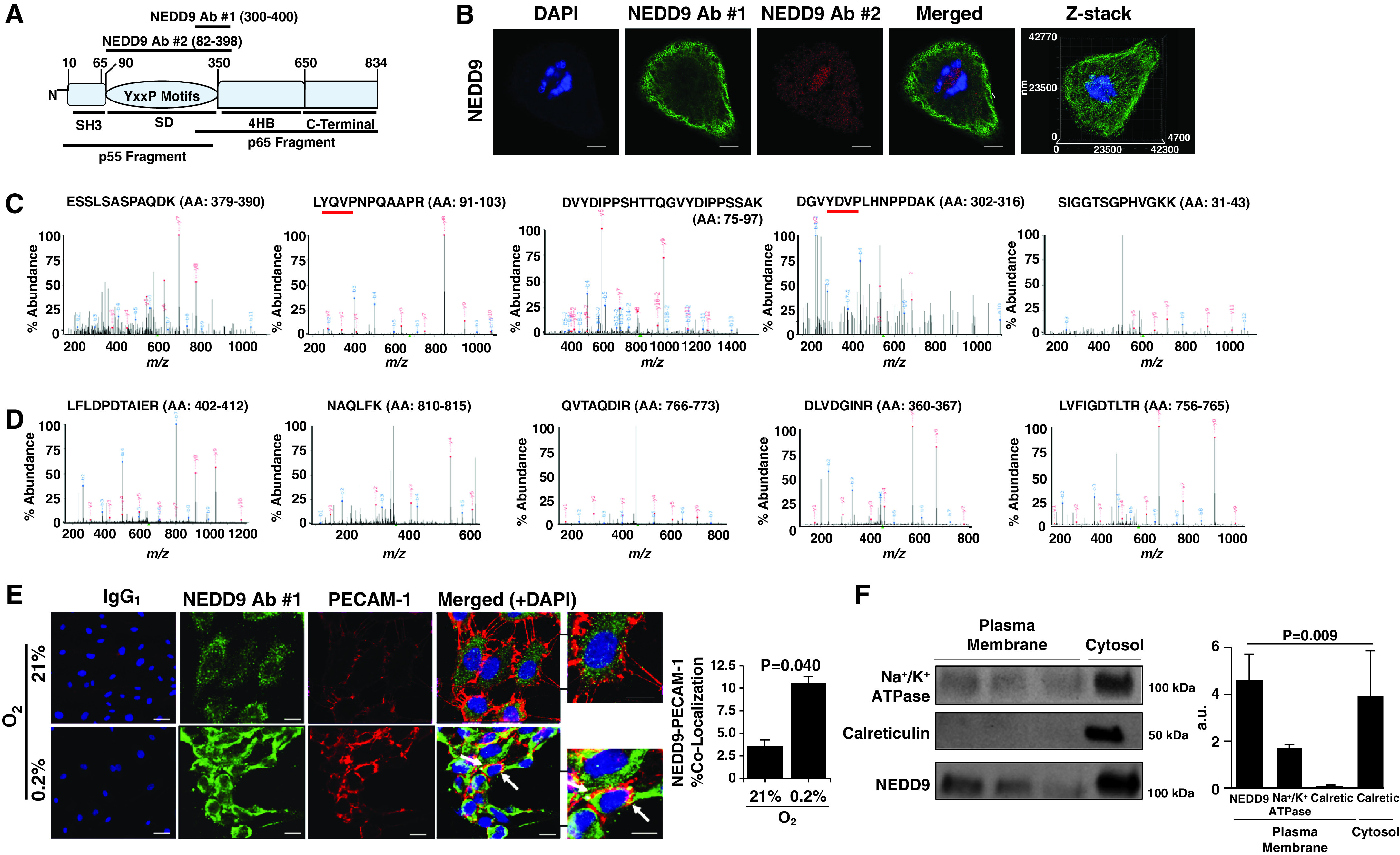
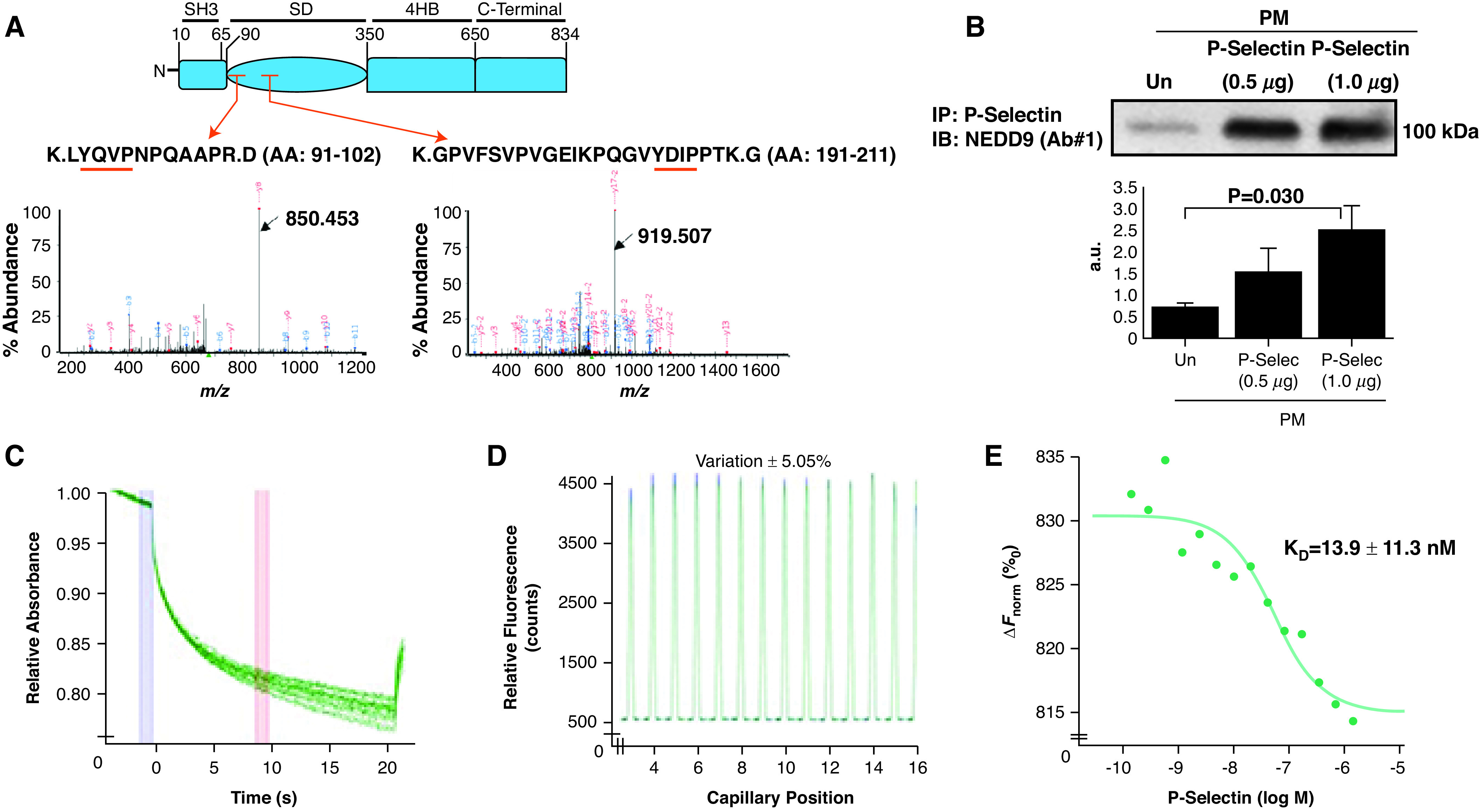
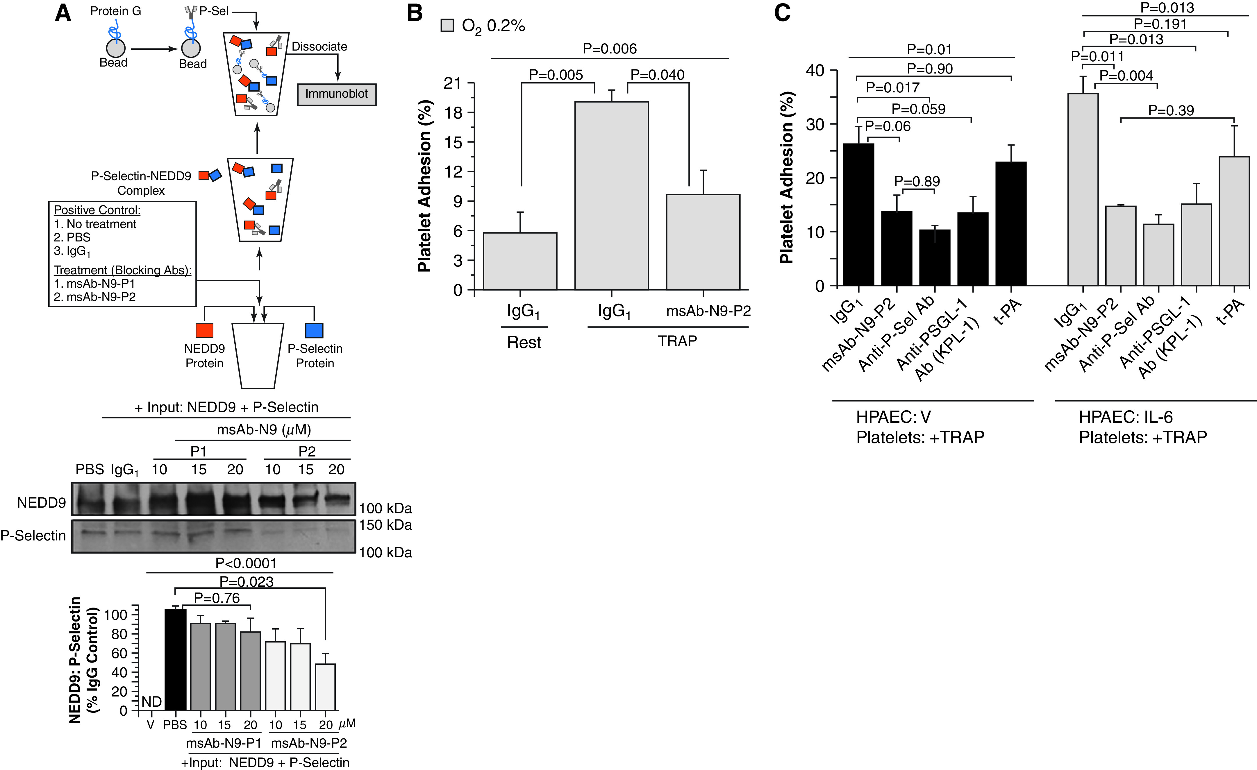
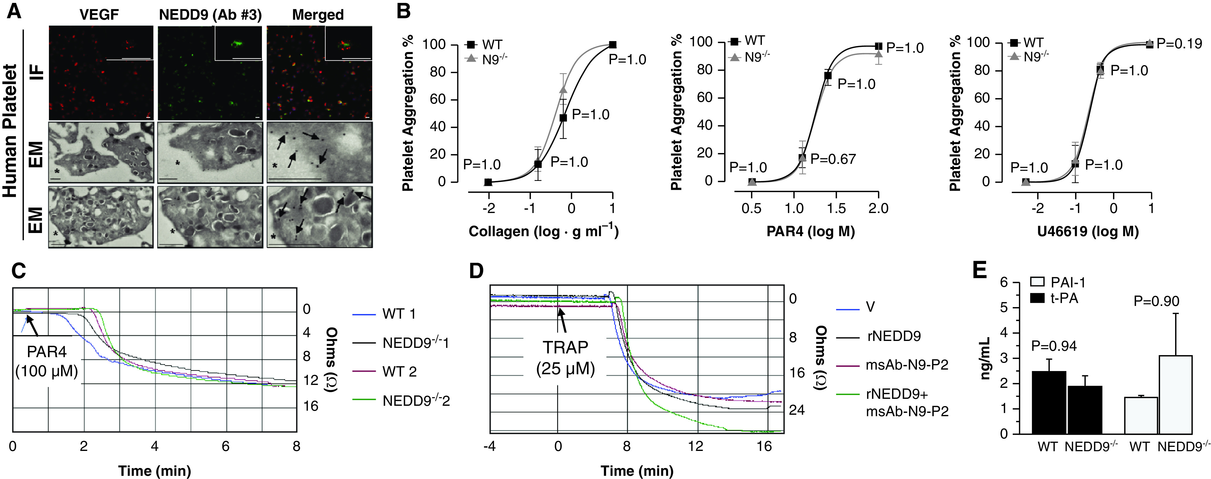
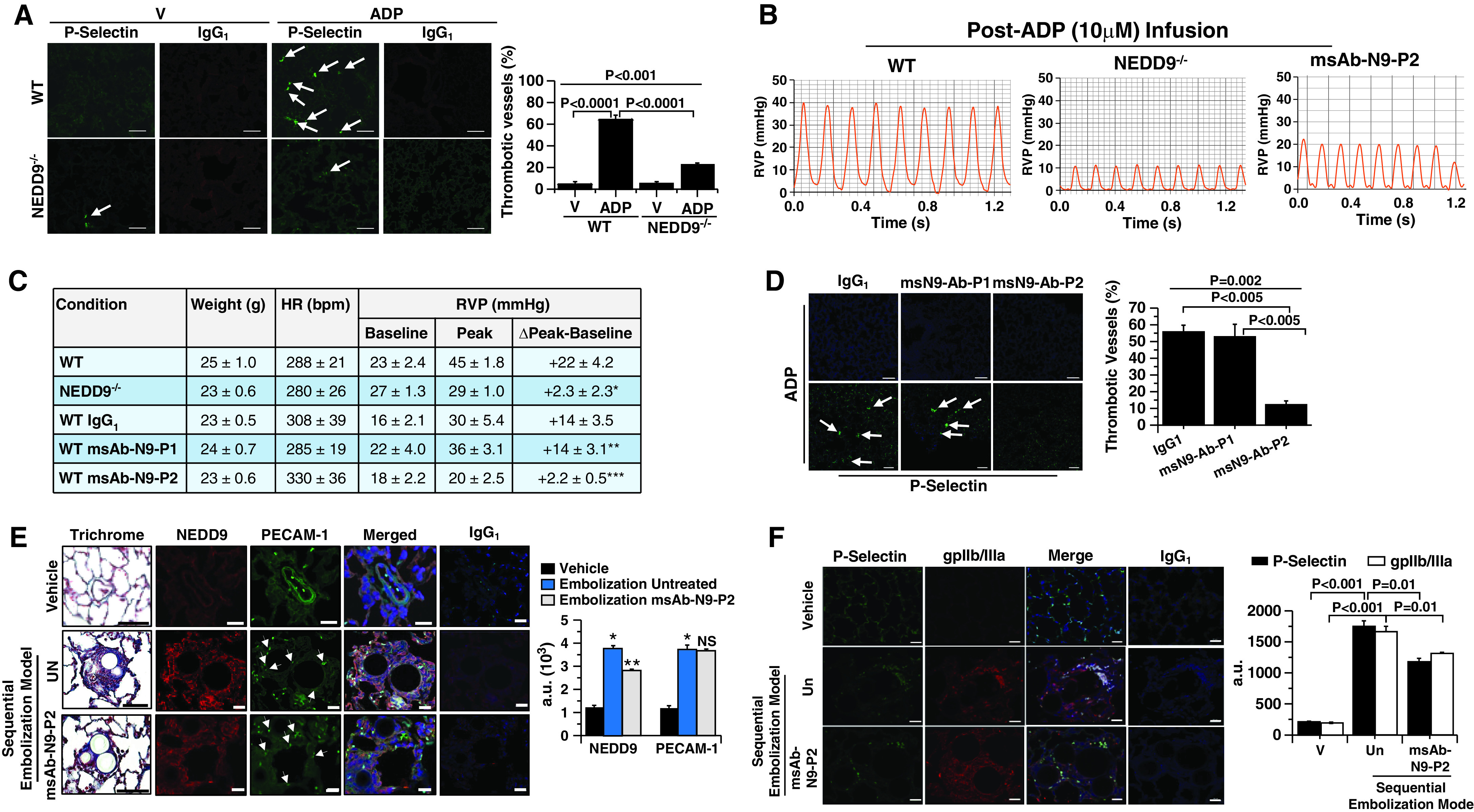
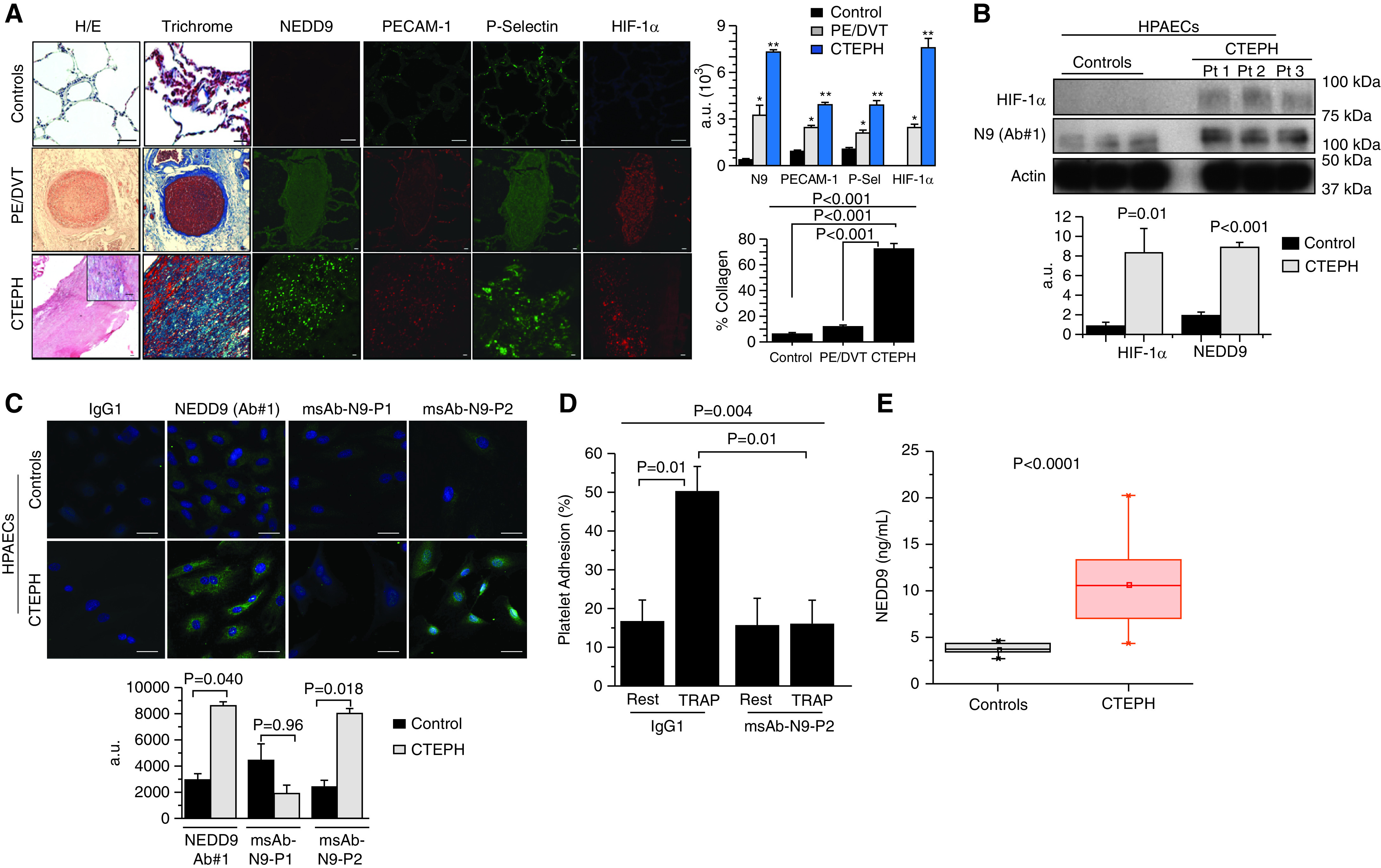
Rationale: Data on the molecular mechanisms that regulate platelet-pulmonary endothelial adhesion under conditions of hypoxia are lacking, but may have important therapeutic implications. Objectives: To identify a hypoxia-sensitive, modifiable mediator of platelet-pulmonary artery endothelial cell adhesion and thrombotic remodeling. Methods: Network medicine was used to profile protein-protein interactions in hypoxia-treated human pulmonary artery endothelial cells. Data from liquid chromatography-mass spectrometry and microscale thermophoresis informed the development of a novel antibody (Ab) to inhibit platelet-endothelial adhesion, which was tested in cells from patients with chronic thromboembolic pulmonary hypertension (CTEPH) and three animal models in vivo. Measurements and Main Results: The protein NEDD9 was identified in the hypoxia thrombosome network in silico. Compared with normoxia, hypoxia (0.2% O2) for 24 hours increased HIF-1α (hypoxia-inducible factor-1α)-dependent NEDD9 upregulation in vitro. Increased NEDD9 was localized to the plasma-membrane surface of cells from control donors and patients with CTEPH. In endarterectomy specimens, NEDD9 colocalized with the platelet surface adhesion molecule P-selectin. Our custom-made anti-NEDD9 Ab targeted the NEDD9-P-selectin interaction and inhibited the adhesion of activated platelets to pulmonary artery endothelial cells from control donors in vitro and from patients with CTEPH ex vivo. Compared with control mice, platelet-pulmonary endothelial aggregates and pulmonary hypertension induced by ADP were decreased in NEDD9-/- mice or wild-type mice treated with the anti-NEDD9 Ab, which also decreased chronic pulmonary thromboembolic remodeling in vivo. Conclusions: The NEDD9-P-selectin protein-protein interaction is a modifiable target with which to inhibit platelet-pulmonary endothelial adhesion and thromboembolic vascular remodeling, with potential therapeutic implications for patients with disorders of increased hypoxia signaling pathways, including CTEPH.

Keywords: hypoxia; platelets; thrombosis.


Figures

Figure 1.
Figure 1.
In human pulmonary artery endothelial cells (HPAECs), the protein NEDD9 is increased by hypoxia–HIF-1α (hypoxia-inducible factor-1α) signaling in vitro and is prothrombotic in silico. (A) Anti-NEDD9 immunoblotting and (B) immunofluorescence were performed on lysates from HPAECs treated with normoxia or hypoxia (10%, 2%, 0.2% O2) for 24 hours (n = 3). IgG1 is the negative control. (C) In contrast to our findings in HPAECs, hypoxia decreased NEDD9 in human brain microvascular endothelial cells (HBMVECs) (n = 3). (D) Inhibition of HIF-1α with siRNA decreases NEDD9 expression in HPAECs treated with hypoxia or cobalt chloride (CoCl2) (250 μmol/L) (n = 3). (E) Venn diagram illustrating the distribution of uniquely differentially expressed gene transcripts analyzed by RNA sequencing in HPAECs and HBMVECs treated with normoxia or hypoxia for 24 hours, and untransfected or transfected with si–HIF-1α (n = 3/condition). The genes related to fibrosis and thrombosis were collected from the curated literature. The gene products (proteins) of HPAEC transcripts that were differentially expressed between normoxia and hypoxia and that were associated with either fibrosis or thrombosis were mapped to the human protein–protein interactome (after Reference 10). NEDD9 (indicated by red oval) was identified in the (F) HPAEC hypoxia fibrosome and the (G) HPAEC hypoxia thrombosome. These findings suggest that NEDD9 is functionally relevant to endophenotypes involved in pulmonary thromboembolism. Scale bars, 20 μm. Data are presented as the mean ± SE. Representative immunoblots and micrographs are shown. a.u. = arbitrary units; Scr = scrambled siRNA (negative) control; si–HIF-1α = HIF-1α siRNA; UN = untreated; V = lipofectamine alone.
Figure 2.
Figure 2.
The NEDD9 substrate domain is expressed on the extracellular plasma membrane of human pulmonary endothelial cells (HPAECs). (A) NEDD9 is a scaffolding protein and in Homo sapiens is composed of 834 amino acids organized in four distinct domains: the SH3, substrate, 4HB, and C-terminal domains. Two NEDD9 cleavage peptide fragments (p55 kD and p65 kD) have been reported previously (22). To determine whether either cleavage product corresponded to differences in NEDD9 localization in HPAECs, anti-NEDD9 immunofluorescence was performed using NEDD9 antibody (Ab) 1 targeting the p55-kD fragment and NEDD9 Ab 2 targeting the p65-kD fragment. (B) Compared with NEDD9 Ab 2, NEDD9 expression detected using NEDD9 Ab 1 was localized predominantly to the perimeter of cells (n = 3). Scale bar = 10 μm. (C) The MS spectra from five abundant peptides detected in trypsin-digested HPAEC lysates immunoprecipitated using NEDD9 Ab 1 corresponded exclusively to the p55-kD fragment, whereas (D) NEDD9 Ab 2 identified NEDD9 peptides corresponding to the p65-kD fragment (n = 3). The red underlining denotes a tyrosine-x-x-proline (YxxP) sequence, where x is another amino acid. (E) Compared with normoxia, hypoxia (0.2% O2) for 24 hours increased colocalization of NEDD9 with the endothelial plasma-membrane protein PECAM-1 (platelet–endothelial cell adhesion molecule 1) analyzed using double immunofluorescence (n = 3) (colocalization indicated by white arrow). IgG1 represents the control. (F) Isolation of HPAEC plasma-membrane fractions was confirmed by Na+/K+ ATPase expression in the absence of (cytosolic) calreticulin, and NEDD9 was analyzed in the plasma-membrane fraction by immunoblotting (n = 5). Scale bar, 40 μm. Data are presented as the mean ± SE. Representative immunoblots and micrographs are presented. a.u. = arbitrary units; Calciretic = calreticulin; MS = mass spectrometry; m/z = mass-to-charge ratio.
Figure 3.
Figure 3.
P-Selec (P-selectin) binds the NEDD9 substrate domain, and NEDD9 modulates platelet–endothelial adhesion. (A) Plasma-membrane fractions incubated with recombinant P-Selec for 1 hour were immunoprecipitated with an anti–P-Selec antibody (Ab). The fragmented ion MS spectra for each of the two detected NEDD9 peptide sequences, both within the substrate domain, are shown (black arrows): K.LYQVPNPQAAPR.D (amino acid [AA], 91–102) (m/z 677.36735 at retention time of 28.1 s) (NEDD9 peptide sequence 1) and K.GPVFSVPVGEIKPQGVYDIPPTK.G (AA, 191–211) (m/z 808.77731 at retention time 35.5 s) (NEDD9 peptide sequence 2) (n = 2 replicates for n = 2 iterations). The red arrows indicate the peptide location within the NEDD9 substrate domain (SD). The red underlining indicates a tyrosine-x-x-proline (YxxP) sequence, where x is another amino acid. (B) Human pulmonary-endothelial-cell plasma-membrane fractions were incubated with exogenous P-Selec, and coimmunoprecipitation was performed using an anti–P-Selec Ab followed by anti–NEDD9 immunoblot NEDD9. Varying concentrations of P-Selec (ligand) (2 μM–0.5 nM) were coincubated with fluorescently labeled NEDD9 (receptor) (20 nM), and microscale thermophoresis was performed to assess macromolecular interactions between these proteins. (C) Raw fluorescence tracings, (D) a capillary scan, and (E) a dose titration curve show a definitive protein–protein interaction between the receptor and ligand (Kd = 13.9 ± 11.3 nM) (n = 2). Data are presented as the mean ± SE. Representative immunoblot and titration curve are shown. a.u. = arbitrary units; ΔFnorm = change in normalized fluorescence; IB = immunoblot; IP = immunoprecipitation; M = molar; MS = mass spectrometry; m/z = mass-to-charge ratio; PM = plasma membrane; Un = untreated.
Figure 4.
Figure 4.
Inhibition of NEDD9 with monospecific polyclonal Ab (msAb)–NEDD9 peptide sequence 2 (N9-P2) decreases NEDD9–P-Sel (P-selectin) complex formation. Anti-NEDD9 antibodies raised against the NEDD9 target of P-Sel were developed in rabbits (msAb–NEDD9 peptide sequence 1 [N9-P1] and msAb–N9-P2). (A) The inhibitory effect of msAb–N9-P1 and msAb–N-P2 on NEDD9–P-Sel complex formation in vitro was analyzed (n = 3). V, a mixture of msAb–N9-P1 and msAb–N9-P2, was incubated without beads as an additional negative control (immunoblot data not shown). (B) In human pulmonary artery endothelial cells (HPAECs) treated with hypoxia (0.2%) for 24 hours, msAb–N9-P2 inhibited TRAP (thrombin receptor agonist peptide) (25 μM)–stimulated platelet–endothelial adhesion significantly (n = 3). IgG1 was the negative control. (C) Under normoxic conditions, HPAECs were treated with IL-6 (25 ng/ml) for 24 hours, and the effect of msAb–N9-P2 (20 μg/ml) on platelet–endothelial adhesion was compared with treatment with anti–P-Sel Ab (anti–P-Sel) (10 μg/ml), PSGL-1 (anti–P-Sel glycoprotein ligand-1 Ab from a KPL-1 clone) (15 μg/ml), and t-PA (tissue plasminogen activator) (15 ng/ml) (n = 3). Similar experiments were performed in untreated HPAECs after stimulation of platelets with TRAP. Data are presented as mean ± SEM. Representative immunoblots are shown. Ab = antibody; ND = not detected; PBS = phosphate-buffered saline; V = vehicle control.
Figure 5.
Figure 5.
N9 (NEDD9) affects platelet–human pulmonary artery endothelial cell adhesion without affecting platelet aggregation. (A) Anti-N9 immunofluorescence (IF) and electron microscopy (EM) immunocytochemistry using N9 antibody (Ab) 3 were performed on platelets isolated from healthy human control donors. For IF, the scale bar was set at 1.5 μm, and the scale bar for the magnified inset was set at 5 μm. For EM, the black arrows identify N9 stain positivity (n = 3); scale bars, 500 nm. (B) No significant difference between wild-type (WT) and N9−/− mice was observed for global platelet aggregation in response to collagen, PAR4 (protease activator receptor) or PAR9, 11-dideoxy-9α, or 11α-methanoepoxy prostaglandin F (U46619) (N = 3 mice/condition). (C) Whole-blood impedance aggregometry was performed in samples acquired from WT and transgenic N9−/− mice stimulated with PAR4 (100 μM) (n = 4). (D) Compared with V control, biologically important differences in impedance aggregometry were not observed in human control blood incubated with rN9 (recombinant N9) (10 ng/ml) or our custom anti-N9 Ab (msAb–N9-P2) (0.08 μg) (n = 3). (E) Plasma expression of t-PA (tissue plasminogen activator) and PAI-1 (plasminogen activator inhibitor-1) in WT or N9−/− mice (n = 4 mice/condition). Data are presented as the mean ± SE. Representative micrographs are shown. M = molar; msAb = monospecific polyclonal Ab; P2 = peptide sequence 2; TRAP = thrombin receptor agonist peptide; V = vehicle; VEGF = vascular endothelial growth factor.
Figure 6.
Figure 6.
The effect of N9 (NEDD9) inhibition with msAb–N9-P2 on platelet–pulmonary endothelial aggregate formation, pulmonary hypertension, and chronic thromboembolic pulmonary arterial remodeling in vivo. (AD) Compared with wild-type (WT) mice, N9−/− mice were resistant to ADP-induced pulmonary arteriolar thrombotic occlusion (arrows) and increased right-ventricular systolic pressure analyzed by anti–P-selectin immunofluorescence and cardiac catheterization, respectively. IgG1 was used as a negative control. Similar effects were observed in WT mice treated with msAb–N9-P2 compared with IgG1 administered 10 minutes before ADP infusion. n = 3–6 mice/condition. For C, *P = 0.005 versus WT, **P = 0.99 versus WT IgG1, and ***P = 0.037 versus WT IgG1; scale bars, 5 μm. Hemodynamic tracings are after administration of maximal ADP dose (10 μM), and full-time course data are presented in Figure E8B. (E) Male and female Wistar rats were randomized to treatment with V (saline) control (n = 3), sequential prothrombotic bead embolization (n = 5), or sequential embolization plus treatment with msAb–N9-P2 (20 μg/ml) (n = 5) (an illustration of the protocol is provided in Figure E6). Paraffin-embedded lung sections were isolated, affected vessels were analyzed for collagen quantity by using Masson trichrome staining, and endothelial N9 and PECAM-1 (platelet–endothelial cell adhesion molecule 1) expression was quantified by using immunofluorescence. Arrows indicate endothelial cells. Scale bars, 20 μm. *P < 0.001, embolization untreated versus vehicle; **P < 0.001 embolization msAb-N9-P2 versus vehicle. (F) Thrombotic remodeling was assessed further by anti–P-selectin and anti-gpIIb/IIIa immunofluorescence. P < 0.001 by ANOVA for P-selectin and P < 0.001 by ANOVA for gpIIb/IIIa. Representative micrographs and hemodynamic tracings are shown. Data are presented as the mean ± SE. Ab = antibody; a.u. = arbitrary units; bpm = beats per minute; gp = glycoprotein; HR = heart rate; ms = monospecific polycolonal; NS = not significant; P1 = peptide sequence 1; P2 = peptide sequence 2; RVP = right ventricular pressure; Un = untreated; V = vehicle.
Figure 7.
Figure 7.
N9 (NEDD9) is increased in chronic thromboembolic pulmonary hypertension (CTEPH). (A) Compared with healthy control donor lung specimens, a step-wise increase was observed in acute pulmonary embolism/deep vein thrombosis (PE/DVT) specimens and CTEPH specimens for vascular fibrosis and HIF-1α (hypoxia-inducible factor-1α), N9, and P-Sel (P-selectin) expression. n = 3–6 samples per condition. Control scale bars, 20 μm; PE/DVT and CTEPH scale bars, 40 μm. *P < 0.01 versus internal control and **P < 0.01 versus PE/DVT. (B) Cultured control human pulmonary artery endothelial cells (HPAECs) and HPAECs from CTEPH specimens were analyzed using anti–HIF-1α and anti-N9 (Ab 1) immunoblotting (n = 6). (C) Anti-N9 immunofluorescence was performed on cultured CTEPH HPAECs using N9 Ab 1, msAb–N9-P1, as a negative control or msAb–N9-P2 (n = 3/condition). IgG1 was used as a negative control. (D) Platelet–endothelial adhesion was analyzed in CTEPH HPAECs and control HPAECs incubated with platelets from healthy donors under basal conditions and after stimulation with TRAP (thrombin receptor agonist peptide) (10 μM) in the presence or absence of msAb–N9-P2 (n = 4). (E) Plasma NEDD9 was increased significantly in patients with CTEPH (n = 27) compared with age-matched and sex-matched healthy controls (n = 7). Scale bars, 20 μm. Representative micrographs and immunoblots are shown. For AD, data are presented as mean ± SE. For E, the means were represented by squares; the medians were represented by horizontal lines; the interquartile ranges were represented by the box distribution; and the maximum and minimum were represented by the y-axis lines. a.u. = arbitrary units; H/E = hematoxylin and eosin; msAb–N9-P1 = monospecific anti-N9 Ab against substrate domain P1; msAb–N9-P2 = monospecific anti-N9 Ab against substrate domain P2; P1 = peptide sequence 1; P2 = peptide sequence 2; Pt = patient.

Comment in

References

    1. Kroll MH, Afshar-Kharghan V. Platelets in pulmonary vascular physiology and pathology. Pulm Circ. 2012;2:291–308. - PMC - PubMed
    1. Rhodes CJ, Im H, Cao A, Hennigs JK, Wang L, Sa S, et al. RNA sequencing analysis detection of a novel pathway of endothelial dysfunction in pulmonary arterial hypertension. Am J Respir Crit Care Med. 2015;192:356–366. - PMC - PubMed
    1. Maron BA, Oldham WM, Chan SY, Vargas SO, Arons E, Zhang YY, et al. Upregulation of steroidogenic acute regulatory protein by hypoxia stimulates aldosterone synthesis in pulmonary artery endothelial cells to promote pulmonary vascular fibrosis. Circulation. 2014;130:168–179. - PMC - PubMed
    1. Kim S-H, Xia D, Kim S-W, Holla V, Menter DG, Dubois RN. Human enhancer of filamentation 1 Is a mediator of hypoxia-inducible factor-1alpha-mediated migration in colorectal carcinoma cells. Cancer Res. 2010;70:4054–4063. - PMC - PubMed
    1. Gay LJ, Felding-Habermann B. Contribution of platelets to tumour metastasis. Nat Rev Cancer. 2011;11:123–134. - PMC - PubMed

Publication types

Substances