. 2021 Jan 1;7(1):eabe1386.
doi: 10.1126/sciadv.abe1386. Print 2021 Jan.

Recessive NOS1AP variants impair actin remodeling and cause glomerulopathy in humans and mice

Affiliations

Recessive NOS1AP variants impair actin remodeling and cause glomerulopathy in humans and mice

Amar J Majmundar et al. Sci Adv. .

Abstract

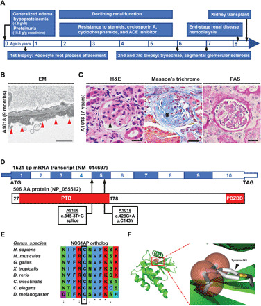
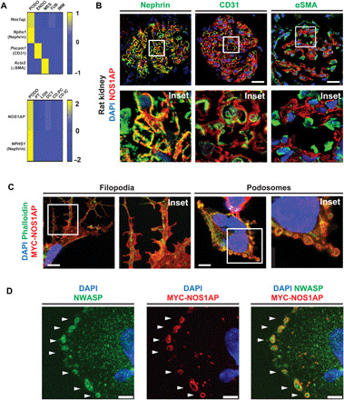
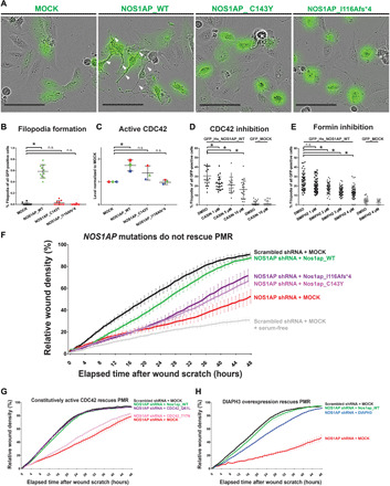
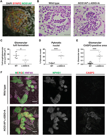
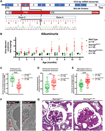
Nephrotic syndrome (NS) is a leading cause of chronic kidney disease. We found recessive NOS1AP variants in two families with early-onset NS by exome sequencing. Overexpression of wild-type (WT) NOS1AP, but not cDNA constructs bearing patient variants, increased active CDC42 and promoted filopodia and podosome formation. Pharmacologic inhibition of CDC42 or its effectors, formin proteins, reduced NOS1AP-induced filopodia formation. NOS1AP knockdown reduced podocyte migration rate (PMR), which was rescued by overexpression of WT Nos1ap but not by constructs bearing patient variants. PMR in NOS1AP knockdown podocytes was also rescued by constitutively active CDC42Q61L or the formin DIAPH3 Modeling a NOS1AP patient variant in knock-in human kidney organoids revealed malformed glomeruli with increased apoptosis. Nos1apEx3-/Ex3- mice recapitulated the human phenotype, exhibiting proteinuria, foot process effacement, and glomerulosclerosis. These findings demonstrate that recessive NOS1AP variants impair CDC42/DIAPH-dependent actin remodeling, cause aberrant organoid glomerulogenesis, and lead to a glomerulopathy in humans and mice.


Figures

Fig. 1
Fig. 1. Homozygous NOS1AP variants in individuals with steroid-resistant NS.
(A) Time bar outlining clinical course of patient A1018 with onset of disease at 4 days of age and progression to end-stage renal disease (ESRD) with subsequent kidney transplant at 8 years of age. (B) Renal biopsy in subject A1018 (9 months) revealed podocyte foot process effacement (red arrowheads) by electron microscopy (EM). Scale bar, 1 μm. (C) Renal biopsy in A1018 (7 years) showed glomerular synechiae formation (arrow) and capillary loop collapse [hematoxylin and eosin (H&E) staining], increased sclerosis (asterisk) (Masson’s trichrome staining), and thickened basement membrane (PAS staining). Scale bar, 50 μM. (D) Coding exon (top bar) and protein domain (bottom bar) structures of NOS1AP are shown with arrows indicating position of variants identified in A5106 and A1018. AA, amino acid. PDZBD, PDZ-binding domain. (E) Evolutionary conservation of primary amino acid sequence shows that cysteine 143 is conserved in NOS1AP orthologs through C. elegans. (F) Modeling of C143Y in the structure of the human NUMB-like protein PTB domain (Protein Data Bank: 3F0W) reveals that, relative to the Cys143 residue in black and gold, the Tyr143 substitution (gray aromatic ring) sterically clashes with neighboring amino acid residues (red spheres) (also see fig. S3H).
Fig. 2
Fig. 2. NOS1AP is expressed by podocytes of mammalian glomeruli and localizes to actin-rich filopodia and podosomes.
(A) NOS1AP mRNA (z-score) was predominantly expressed by podocytes from single-cell mRNA sequencing data (6, 40). Top: Nos1ap expression was highest in the podocyte cluster (Nphs1 and nephrin) relative to the endothelial cell cluster (Pecam1 and CD31), mesangial cell cluster (Acta2 and α-smooth muscle actin), and renal tubular and immune cells in murine glomeruli (6). Bottom: NOS1AP expression is similarly highest in the podocyte cluster relative to other nephron epithelial cell clusters from adult human kidney (40). PODO, podocytes; ENDO, endothelial cells; MES, mesangial cells; TUB, tubular epithelial cells; IMM, immune cells; PT, proximal tubular epithelial cells; LOH, Loop of Henle epithelial cells; DCT, distal convoluted tubular epithelial cells; CD:PC, collecting duct principal cells; CD:IC, collecting duct intercalated cells. (B) IF confocal microscopy imaging of rat kidney sections demonstrates NOS1AP colocalization (yellow) with podocyte slit diaphragm marker nephrin but not endothelial cell marker CD31 or mesangial cell marker αSMA. Insets are shown below each image. Scale bars, 25 μm. (C) In a human podocyte cell line, overexpression of MYC-tagged WT NOS1AP (MYC-NOS1AP) induced the formation of filopodia (left column) and peripheral ring structures (right column) in which NOS1AP colocalized with F-actin. (Images 26 hours after transfection.) Scale bars, 7.5 μm. (D) MYC-NOS1AP overexpression in a human podocyte cell line identifies peripheral ring structures as podosomes by colocalization of NOS1AP with NWASP (white arrows). Scale bars, 5 μm.
Fig. 3
Fig. 3. NOS1AP induces filopodia formation and is essential for PMR, potentially via a CDC42/DIAPH pathway.
(A) Podocytes were transfected with GFP-plasmids (MOCK, WT NOS1AP, and NOS1AP patient mutation constructs). Representative images are shown with white arrows pointing to filopodia. Scale bars, 100 μm. (B) WT NOS1AP increased filopodia formation to 59% (range, 40 to 78%). NS mutant construct overexpression (C143Y and I116Afs*4) failed to induce filopodia (3 and 0%). [One-way analysis of variance (ANOVA), *P < 0.05; n.s., nonsignificant]. (C) Human embryonic kidney (HEK) 293T cells were transfected with NOS1AP WT or cDNA reflecting patient variants. WT NOS1AP overexpression caused a significant increase in active CDC42 levels, while NS patient variant constructs did not (one-way ANOVA; unique colors for independent experiments). (D) CDC42 inhibitor CASIN blocked filopodia formation in NOS1AP-transfected podocytes (one-way ANOVA, see fig. S6B for representative images). (E) Formin inhibitor SMIFH2 attenuated filopodia formation in NOS1AP-transfected podocytes (one-way ANOVA, see fig. S6C for representative images). DMSO, dimethyl sulfoxide. (F) Knockdown of NOS1AP (red) resulted in reduced PMR compared with scrambled shRNA control cells (black), which was rescued by overexpression of WT Nos1ap (green) but not NS patient variant constructs (purple and pink). (G) Reduced PMR upon knockdown of NOS1AP (red) was rescued by overexpression of WT Nos1ap (green) and a constitutively active human CDC42Q61L construct (purple) but not the hypomorphic CDC42T17N construct (pink). (H) Reduced PMR upon knockdown of NOS1AP (red) was rescued by overexpression of WT Nos1ap (green) and partially rescued by the formin DIAPH3 (blue).
Fig. 4
Fig. 4. Kidney organoids bearing the homozygous NOS1AP patient variant c.428G>A exhibit aberrantly formed glomeruli.
(A) IF imaging demonstrated NOS1AP localization to podocytes in organoid glomeruli adjacent to the podocyte marker SYNPO. Scale bar, 20 μm. (B) In WT and NOS1AP c.428G>A mutant organoids (PAS staining), glomerular tufts (within white dashed lines) were defined as linear podocyte monolayers organized bilaterally about established extracellular matrix (black lines) and were reduced in NOS1AP mutant organoids. NOS1AP mutant glomeruli also exhibited increased pyknotic nuclei (arrowheads), indicative of cell death. Scale bars, 20 μm. Also see fig. S8H for additional images of PAS staining. Scale bars, 20 μm. (C) Cumulative glomerular tuft length measurement is plotted from two to three independent fields (dots) from three independent organoid cultures. NOS1AP mutant organoids demonstrate significantly lower cumulative tuft length than WT organoids (Mann-Whitney U test, *P < 0.05). (D) Percentage of pyknotic nuclei is plotted from independent fields and organoids as in (B). NOS1AP mutant organoids exhibit significantly increased pyknotic nuclei relative to WT organoids (Mann-Whitney U test, ***P < 0.001). (E) Quantification of active caspase-3 (CASP3) staining in glomerular regions is shown from three paired differentiation experiments to substantiate increased cell death that was indicated by pyknotic nuclei in (B) and (D). NOS1AP mutant organoids demonstrate elevated apoptosis (Mann-Whitney U test, ***P < 0.001). (F) Whole mount IF of organoids for apoptotic marker cleaved CASP3 is shown. CASP3 staining is increased in glomeruli (NPHS1) of NOS1AP mutant organoid glomeruli, relative to WT organoids. CASP3 signal in tubular segments (HNF4A) is not increased. Scale bars, 100 μm. Also see fig. S8G for CASP3 staining in organoids derived from the second independent iPSC cell line PCS201010.
Fig. 5
Fig. 5. Nos1apEx3−/Ex3− mice develop glomerular proteinuria, foot process effacement, and mesangial matrix expansion.
(A) Coding exon (top) and protein domain (bottom) structures of murine Nos1ap and Sanger sequencing trace for Nos1apEx3−/Ex3− cDNA are shown. Exon 3 deletion (red rectangle) causes an in-frame deletion within the PTB domain. (B) Urinary albumin/creatinine ratios were measured. Nos1apEx3−/Ex3− mice develop significant albuminuria. (C) Podocyte foot process density was quantified in transmission electron microscopy (TEM) images for Nos1apEx3−/+ and Nos1apEx3−/Ex3− mice [five animals per genotype, 11 months old (three) and 16 months old (two); 65 and 69 capillary loops per genotype, respectively]. Foot process effacement is observed in homozygous Nos1apEx3−/Ex3− mice. (Each dot represents one capillary loop, and bars represent minimum to maximum; Kruskal-Wallis test, ***P < 0.001). (D) Increased glomerular basement membrane (GBM) thickness was observed in TEM images of homozygous Nos1apEx3−/Ex3− mice. (Bars represent minimum to maximum; Kruskal-Wallis test, **P < 0.01). (E) Mesangial matrix area per glomerulus in PAS staining was increased in homozygous Nos1apEx3−/Ex3− mice. [Each dot represents one glomerulus, and bars represent minimum to maximum; five animals per genotype, 11 months old (three) and 16 months old (two); 158 and 179 images per genotype, respectively; Kruskal-Wallis test, ***P < 0.001]. (F) Representative glomerular TEM images for Nos1apEx3−/+ and Nos1apEx3−/Ex3− mice demonstrate podocyte foot process effacement and thickened basement membranes. Scale bars, 1 μm. (G) Representative PAS images show mesangial matrix expansion, partially collapsed capillary loops, and thickened basement membranes in Nos1apEx3−/Ex3− mice. Scale bars, 200 μm. Also see fig. S10.

References

    1. W. Harmon, R. Fine, S. Alexander, B. Warady, M. Benfield, S. Goldstein, R. McDonald, K. Martz, D. Stablein, NAPRTCS 2008 Annual Report (2008).
    1. Wiggins R.-C., The spectrum of podocytopathies: A unifying view of glomerular diseases. Kidney Int. 71, 1205–1214 (2007). - PubMed
    1. Connaughton D. M., Kennedy C., Shril S., Mann N., Murray S. L., Williams P. A., Conlon E., Nakayama M., van der Ven A. T., Ityel H., Kause F., Kolvenbach C. M., Dai R., Vivante A., Braun D. A., Schneider R., Kitzler T. M., Moloney B., Moran C. P., Smyth J. S., Kennedy A., Benson K., Stapleton C., Denton M., Magee C., O’Seaghdha C. M., Plant W. D., Griffin M. D., Awan A., Sweeney C., Mane S. M., Lifton R. P., Griffin B., Leavey S., Casserly L., de Freitas D. G., Holian J., Dorman A., Doyle B., Lavin P. J., Little M. A., Conlon P. J., Hildebrandt F., Monogenic causes of chronic kidney disease in adults. Kidney Int. 95, 914–928 (2019). - PMC - PubMed
    1. Sadowski C. E., Lovric S., Ashraf S., Pabst W. L., Gee H. Y., Kohl S., Engelmann S., Vega-Warner V., Fang H., Halbritter J., Somers M. J., Tan W., Shril S., Fessi I., Lifton R. P., Bockenhauer D., El-Desoky S., Kari J. A., Zenker M., Kemper M. J., Mueller D., Fathy H. M., Soliman N. A.; SRNS Study Group and Friedhelm Hildebrandt , A single-gene cause in 29.5% of cases of steroid-resistant nephrotic syndrome. J. Am. Soc. Nephrol. 26, 1279–1289 (2015). - PMC - PubMed
    1. Warejko J. K., Tan W., Daga A., Schapiro D., Lawson J. A., Shril S., Lovric S., Ashraf S., Rao J., Hermle T., Jobst-Schwan T., Widmeier E., Majmundar A. J., Schneider R., Gee H. Y., Schmidt J. M., Vivante A., van der Ven A. T., Ityel H., Chen J., Sadowski C. E., Kohl S., Pabst W. L., Nakayama M., Somers M. J. G., Rodig N. M., Daouk G., Baum M., Stein D. R., Ferguson M. A., Traum A. Z., Soliman N. A., Kari J. A., Desoky S. E., Fathy H., Zenker M., Bakkaloglu S. A., Müller D., Noyan A., Ozaltin F., Cadnapaphornchai M. A., Hashmi S., Hopcian J., Kopp J. B., Benador N., Bockenhauer D., Bogdanovic R., Stajić N., Chernin G., Ettenger R., Fehrenbach H., Kemper M., Munarriz R. L., Podracka L., Büscher R., Serdaroglu E., Tasic V., Mane S., Lifton R. P., Braun D. A., Hildebrandt F., Whole exome sequencing of patients with steroid-resistant nephrotic syndrome. Clin. J. Am. Soc. Nephrol. 13, 53–62 (2018). - PMC - PubMed

Publication types

LinkOut - more resources