. 2021 Jan 15;7(3):eabe0726.
doi: 10.1126/sciadv.abe0726. Print 2021 Jan.

Strontium ions protect hearts against myocardial ischemia/reperfusion injury

Affiliations

Strontium ions protect hearts against myocardial ischemia/reperfusion injury

Min Xing et al. Sci Adv. .

Abstract

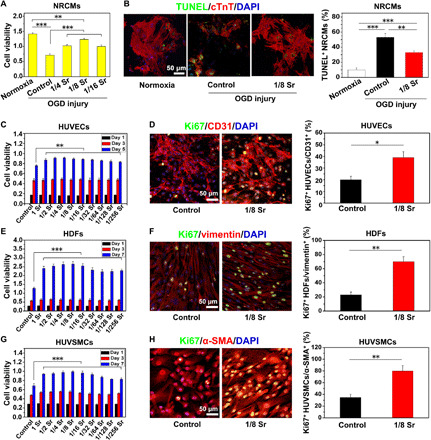
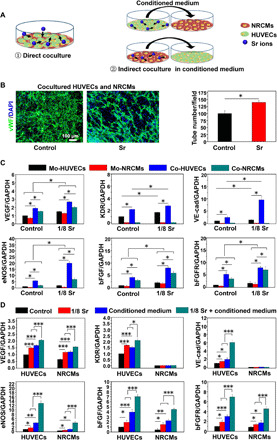
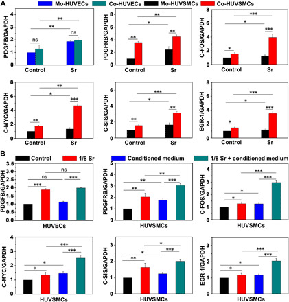
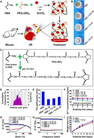
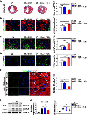
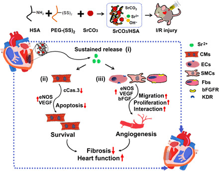
Timely restoration of blood supply following myocardial infarction is critical to save the infarcted myocardium, while reperfusion would cause additional damage. Strontium ions have been shown to promote angiogenesis, but it is unknown whether they can save the damaged myocardium. We report that myocardial ischemia/reperfusion (I/R)-induced functional deterioration and scar formation were notably attenuated by injection of strontium ion-containing composite hydrogels into murine infarcted myocardium at 20 minutes of reperfusion following 60 minutes of ischemia. These beneficial effects were accompanied by reduced cardiomyocyte apoptosis and increased angiogenesis. The effects of strontium ions were further confirmed by the enhanced viability of cardiomyocytes and stimulated angiogenesis in vitro. These findings are the first to reveal the cardioprotective effects of strontium ions against I/R injury, which may provide a new therapeutic approach to ischemic heart disease at a lower cost, with higher stability, and with potentially greater safety.


Figures

Fig. 1
Fig. 1. Sr ions reduce NRCM apoptosis after OGD injury and promote blood vessel–related cell proliferation.
(A) The viability of NRCMs measured by Cell Counting Kit-8 (CCK8) in the medium supplemented with different concentrations of Sr ion after OGD injury. (B) TUNEL staining (green), cTnT staining (red), and DAPI (4′,6-diamidino-2-phenylindole) staining (blue) in NRCMs after OGD injury and quantitative analysis of TUNEL+ NRCMs (10 pictures for each group). The corresponding concentrations of Sr ion with the 1/4 to 1/16 dilution ratio for NRCM culture are shown in table S1. (C to H) The cell viability and proliferation of human umbilical vein endothelial cells (HUVECs) (C and D), human dermal fibroblasts (HDFs) (E and F), and human umbilical vein smooth muscle cells (HUVSMCs) (G and H) after culturing in the medium supplemented with Sr ions at different concentrations. They were respectively revealed by CCK8 and the immunofluorescence of Ki67, followed by quantitative analysis of Ki67+ after Sr ion treatment for 5 days (HUVECs) or 7 days (HDFs and HUVSMCs). The corresponding concentrations of Sr ion with the dilution ratio of 1 to 1/256 for HUVEC, HDF, and HUVSMC culture are respectively shown in table S1. Experiments were conducted in triplicate. All data are presented as means ± SEM. An unpaired t test was used to compare between any two groups. One-way analysis of variance (ANOVA) was used to compare between three or more groups. *P < 0.05, **P < 0.01, and ***P < 0.001.
Fig. 2
Fig. 2. Sr ions stimulate paracrine-mediated angiogenic effects of HUVECs and NRCMs after 3 days of coculture.
(A) Schematic of direct and indirect coculture of HUVECs and NRCMs. (B) Images and quantitative analysis of von Willebrand factor (vWF) staining for the tube formation ability of cocultured HUVECs with control medium and Sr ion–containing medium (10 pictures for each group). An unpaired t test was used for statistical analyses. *P < 0.05. (C) qRT-PCR analysis of the expression levels of angiogenesis-related genes [VEGF, KDR, vascular endothelial–cadherin (VE-cad), eNOS, bFGF, and bFGFR] in HUVECs and NRCMs that were acquired from monocultured cells or cocultured cells in each treatment group. Experiments were conducted in triplicate. An unpaired t test was used for statistical analyses. All data are presented as means ± SEM. *P < 0.05 was considered statistically significant. Mo, monocultured; Co, cocultured. (D) qRT-PCR analysis of the angiogenic gene (VEGF, KDR, VE-cad, eNOS, bFGF, and bFGFR) expression in HUVECs treated with normal medium and conditioned medium from NRCMs, which were treated with Sr ions or not, as well as in NRCMs treated with normal medium and conditioned medium from HUVECs, which were treated with Sr ions or not. Experiments were conducted in triplicate. One-way ANOVA was used for statistical analyses. All data are presented as means ± SEM. *P < 0.05, **P < 0.01, and ***P < 0.001.
Fig. 3
Fig. 3. Sr ions stimulate paracrine-mediated proliferation effects of HUVSMCs after 3 days of coculture with HUVECs.
(A) qRT-PCR analysis of the expression levels of PDGFB in monocultured HUVECs and cocultured HUVECs and gene expression of PDGFRB, C-FOS, C-MYC, C-SIS, and EGR-1 in monocultured HUVSMCs and cocultured HUVSMCs in each treatment group. An unpaired t test was used for statistical analyses. (B) qRT-PCR analysis of the expression levels of PDGFB in HUVECs treated with conditioned medium from HUVSMCs with or without Sr ions and gene expression of PDGFRB, C-FOS, C-MYC, C-SIS, and EGR-1 in HUVSMCs treated with conditioned medium from HUVECs with or without Sr ions. Experiments were conducted in triplicate; one-way ANOVA was used for statistical analyses. All data are presented as means ± SEM. *P < 0.05, **P < 0.01, and ***P < 0.001. ns, no statistical significance.
Fig. 4
Fig. 4. Construct of injectable SrCO3/HSA composite hydrogels (HSA).
(A) Design schematic of SrCO3/HSA composite hydrogels for injection into I/R mice. (B) The pictures of SrCO3/HSA composite hydrogels with different SrCO3 contents. (C) The gelation mechanism and chemical reactions between HSA and PEG-(SS)2. (D) The particle size distribution of the SrCO3 powder. (E) Gelling time of SrCO3/HSA composite hydrogels. (F) Concentrations of Sr ions released from hydrogels. Experiments were conducted in triplicate. (G) Elastic moduli (G′) and viscous moduli (G″) as a function of the strain of HSA hydrogel and HSA + 1% Sr composite hydrogel. (H) G′ and G″ as a function of the frequency of HSA hydrogel and HSA + 1% Sr composite hydrogel. (I) The weight loss of the HSA hydrogel, HSA + 1% Sr composite hydrogel, and SrCO3 powders inside the HSA + 1% Sr composite hydrogel after soaking in tris-HCl buffer solution (pH 7.4) at different time points. Experiments were conducted in triplicate. All data are presented as means ± SEM. (Photo credit: Min Xing, State Key Laboratory of High Performance Ceramics and Superfine Microstructure, Shanghai Institute of Ceramics, Chinese Academy of Sciences, Shanghai 200050, P. R. China.)
Fig. 5
Fig. 5. SrCO3/HSA hydrogels improve the recovery of mice cardiac function after I/R.
(A) Schematic of I/R model establishment, hydrogel injection, and echocardiography (ECO) analysis. (B) Kaplan-Meier survival curves of mice during the 28 days after I/R. (C) Body weights of mice after I/R. (D) Representative echocardiograms obtained from the mid-papillary muscle region of the left ventricle of mice at day 28 (D28) after I/R. (E) Cardiac function measured by the percentage of LVEF and LVFS at days 1 and 28 after I/R. n = 10 each. All data are presented as means ± SEM. Comparisons between three groups were performed using two-way ANOVA. **P < 0.01.
Fig. 6
Fig. 6. SrCO3/HSA hydrogels inhibit myocardial fibrosis, increase angiogenesis, and reduce cardiomyocyte apoptosis.
(A) Representative cross-sectional images and quantitative data of scar area in LV on 5-μm slices stained with Masson’s trichrome at day 28 after I/R. n = 5 hearts for each group. (B to D) Images and quantification of α-SMA+ blood vessels (B), CD31+ cells (C), and VEGF (D) in the border zone of infarcted hearts 28 days after I/R. Scale bars, 50 μm. n = 4 hearts for each group. HPF, high-power field. (E) Representative and quantification of staining for TUNEL+ cardiomyocytes (CMs) in the border zone of infarcted hearts at day 3 after I/R. Scale bars, 50 μm. n = 5 to 6 hearts for each group. (F and G) Representative and averaged Western blot analysis for cleaved caspase-3 (cCas.3) and glyceraldehyde 3-phosphate dehydrogenase (GAPDH) in LV heart tissues at day 3 after I/R. n = 4 hearts each. (H) The concentrations of cardiac injury marker LDH of the mice serum at day 3 after I/R. n = 4 to 6 hearts each. Ctrl, I/R control; HSA, I/R + HSA; 1% Sr, I/R + HSA + 1% Sr. All values are expressed as means ± SEM; one-way ANOVA was used for statistical analyses. *P < 0.05 and ***P < 0.001.
Fig. 7
Fig. 7. The schematic diagram of the SrCO3/HSA hydrogels in myocardium repair after I/R.
Various underlying mechanisms contributed to myocardial repair for SrCO3/HSA hydrogels after I/R: (i) SrCO3/HSA hydrogels sustain the release of Sr ions in the infarct heart; (ii) Sr ions inhibit the apoptosis of CMs through reducing the activity of caspase-3; (iii) Sr ions promote angiogenesis via increasing proliferation, strengthening paracrine capacity, and stimulating the interaction of cardiac cells. Ultimately, alleviative fibrosis and elevated cardiac function are achieved after the treatment of SrCO3/HSA hydrogels. ECs, endothelium cells; SMCs, smooth muscle cells; Fbs, fibroblasts.

References

    1. Zarrinkoub R., Wettermark B., Wändell P., Mejhert M., Szulkin R., Ljunggren G., Kahan T., The epidemiology of heart failure, based on data for 2.1 million inhabitants in Sweden. Eur. J. Heart Fail. 15, 995–1002 (2013). - PubMed
    1. Caccioppo A., Franchin L., Grosso A., Angelini F., D’Ascenzo F., Brizzi M. F., Ischemia reperfusion injury: Mechanisms of damage/protection and novel strategies for cardiac recovery/regeneration. Int. J. Mol. Sci. 20, 5024 (2019). - PMC - PubMed
    1. Vinten-Johansen J., Zhao Z.-Q., Jiang R., Zatta A. J., Dobson G. P., Preconditioning and postconditioning: Innate cardioprotection from ischemia-reperfusion injury. J. Appl. Physiol. 103, 1441–1448 (2007). - PubMed
    1. Davidson S. M., Ferdinandy P., Andreadou I., Bøtker H. E., Heusch G., Ibáñez B., Ovize M., Schulz R., Yellon D. M., Hausenloy D. J., Garcia-Dorado D.; CARDIOPROTECTION COST Action (CA16225) , Multitarget strategies to reduce myocardial ischemia/reperfusion injury: JACC review topic of the week. J. Am. Coll. Cardiol. 73, 89–99 (2019). - PubMed
    1. Zhao Z. Q., Corvera J. S., Halkos M. E., Kerendi F., Wang N. P., Guyton R. A., Vinten-Johansen J., Inhibition of myocardial injury by ischemic postconditioning during reperfusion: Comparison with ischemic preconditioning. Am. J. Physiol. Heart Circ. Physiol. 285, H579–H588 (2003). - PubMed

Publication types