. 2021 Jan 20;7(4):eabe2116.
doi: 10.1126/sciadv.abe2116. Print 2021 Jan.

Linkage-specific deubiquitylation by OTUD5 defines an embryonic pathway intolerant to genomic variation

Affiliations

Linkage-specific deubiquitylation by OTUD5 defines an embryonic pathway intolerant to genomic variation

David B Beck et al. Sci Adv. .

Abstract

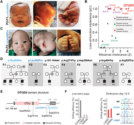
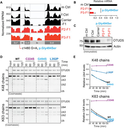
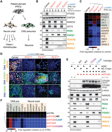
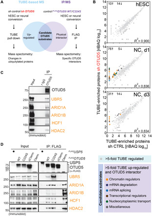
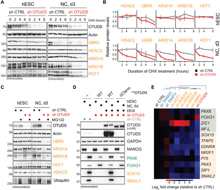
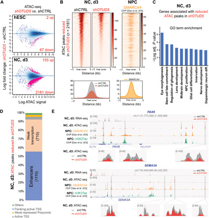
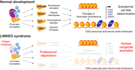
Reversible modification of proteins with linkage-specific ubiquitin chains is critical for intracellular signaling. Information on physiological roles and underlying mechanisms of particular ubiquitin linkages during human development are limited. Here, relying on genomic constraint scores, we identify 10 patients with multiple congenital anomalies caused by hemizygous variants in OTUD5, encoding a K48/K63 linkage-specific deubiquitylase. By studying these mutations, we find that OTUD5 controls neuroectodermal differentiation through cleaving K48-linked ubiquitin chains to counteract degradation of select chromatin regulators (e.g., ARID1A/B, histone deacetylase 2, and HCF1), mutations of which underlie diseases that exhibit phenotypic overlap with OTUD5 patients. Loss of OTUD5 during differentiation leads to less accessible chromatin at neuroectodermal enhancers and aberrant gene expression. Our study describes a previously unidentified disorder we name LINKED (LINKage-specific deubiquitylation deficiency-induced Embryonic Defects) syndrome and reveals linkage-specific ubiquitin cleavage from chromatin remodelers as an essential signaling mode that coordinates chromatin remodeling during embryogenesis.


Figures

Fig. 1
Fig. 1. Hemizygous variants in OTUD5 cause multiple congenital anomaly disorder.
(A) Clinical photos showing craniofacial (retrognathia, midface hypoplasia, hypertelorism, low-set posteriorly rotated ears, and craniosynostosis) and digital anomalies (bilateral postaxial polydactyly of the hands and feet) of patient P2 carrying the p.Gly494Ser variant of OTUD5. Photo credit: With permission of the subjects’ legal guardian. (B) Among several OTU DUBs, OTUD5 is the strongest candidate for hypomorphic mutations leading to disease given high loss-of-function and missense intolerance scores. Loss-of-function intolerance (pLI) and missense intolerance (Z) were determined for all OTU DUBs using gnomAD. (C) Clinical photos showing craniofacial (retrognathia, midface hypoplasia, hypertelorism, low-set posteriorly rotated ears, and craniosynostosis) and digital anomalies (bilateral postaxial polydactyly of the hands and feet) of patient P4 carrying the p.Leu352Pro variant of OTUD5. (D) Genetic pedigrees of 10 patients from seven families with hemizygous variants in OTUD5, all with overlapping phenotypes. (E) Domain structure of OTUD5 indicating the location of the patient mutations. Variants associated with the most severe phenotypes, p.Gly494Ser and p.Leu352Pro, are highlighted in blue colors. NLS, nuclear localization sequence. (F) CRISPR-mediated knockout of OTUD5 or knock-in of the p.Gly494Ser or p.Leu352Pro patient variants results in embryonic lethality. Left: Mouse zygotes were injected with Cas9 complexed with gRNAs and respective repair oligos and transferred into pseudo-pregnant recipient mice. Percentage of live-born pups with edited alleles (knockout or knock-in) for a nonessential gene (control), OTUD5L352P, or OTUD5G494S is shown (n > 70 injected embryos per condition). Right: Mouse embryos were injected with gRNA-loaded Cas9 and respective repair oligos and implanted into mice. Pregnant mice were euthanized and embryos were isolated at E12.5. Percentage of pups with edited alleles (knockout or knock-in) for OTUD5L352P or OTUD5G494S injections are shown (n > 70 injected embryos per condition). Sanger sequencing depicting examples of E12.5 knock-in embryos is shown.
Fig. 2
Fig. 2. OTUD5 patient mutations are hypomorphic and reduce OTUD5 levels or K48-ubiuqitin cleavage activity.
(A) The c.1480 G>A, p.Gly494Ser mutation is located in a 5′ splice site and leads to intron retention and reduction of OTUD5 mRNA levels as revealed by RNA-seq of patient-derived fibroblasts. RNA-seq reads were differentially scaled to visualize intron retention. m Ctrl, male control; f Ctrl, female control; mother carrier, mother F1. (B) The c.1480 G>A p.Gly494Ser mutation results in a decrease in OTUD5 mRNA levels in patient-derived fibroblasts as determined by qRT-PCR (n = 3 biological replicates, error bars denote SD). (C) The p.Gly494Ser mutation results in a decrease in OTUD5 protein levels as revealed by immunoblotting of lysates of patient-derived fibroblasts using indicated antibodies. (D) The Leu352Pro mutation specifically reduces OTUD5’s K48-ubiquitin chain cleavage activity. WT FLAGHAOTUD5 (WT), catalytically inactive FLAGHAOTUD5 (C224S), and patient variant FLAGHAOTUD5 (G494S and L352P) were purified from HEK 293T cells and incubated with K48- or K63-tetra ubiquitin chains for the indicated time periods and analyzed by colloidal Coomassie-stained SDS–polyacrylamide gel electrophoresis gels. (E) Quantification of three independent in vitro deubiquitylation experiments as shown in (D) (error bars denote SEM). Intensity of Ub4 band is relative to the sum of intensity of Ub3, Ub2, and Ub bands. RPKM, reads per kilobase per million mapped reads.
Fig. 3
Fig. 3. OTUD5 regulates CNS precursor and neural crest cell differentiation via its K48-ubiquitin chain–specific deubiquitylation activity.
(A) Schematic overview of the neural conversion paradigm. (B) Reduction of OTUD5 levels causes aberrant neural conversion. iPSCs derived from OTUD5 p.Gly494Ser patients or the maternal carrier were subjected to neural conversion for 6 days. Differentiation was monitored by immunoblotting using indicated antibodies against hESC, CNS precursor, and neural crest markers. (C) Same experimental approach as described in (B), but cells were analyzed by qRT-PCR for expression of CNS precursor markers (green) and neural crest markers (orange). Marker expression was normalized to carrier control followed by hierarchical clustering. RPL27, endogenous control. (D) Same experimental approach as described in (B), but cells were subjected to neural conversion for 9 days and analyzed by immunofluorescence microscopy using antibodies against indicated CNS precursor and neuronal markers (green) or neural crest markers (orange). Scale bars, 20 μm. (E) K48-ubiquitin chain–specific deubiquitylation activity of OTUD5 is required for proper CNS precursor and neural crest differentiation. hES H1 cells stably expressing shRNA-resistant and doxycycline-inducible WT, catalytically inactive (C224S), or K48 chain cleavage–deficient (L352P) HAOTUD5 were generated. Cells were depleted of endogenous OTUD5 using shRNA as indicated, treated with or without doxycycline (DOX), and subjected to neural conversion (NC) for 6 days. This was followed by immunoblotting using the indicated antibodies against hESC, CNS precursor, and neural crest markers. (F) Same experimental approach as described in (E), but cells were analyzed by qRT-PCR analysis for expression of CNS precursor markers (green) and neural crest markers (orange). Marker expression was normalized to shcontrol followed by hierarchical cluster analysis. RPL27, endogenous control.
Fig. 4
Fig. 4. Identification of chromatin remodelers as candidate substrates of OTUD5.
(A) Strategy used to isolate high-probability substrates of OTUD5. Two independent proteomic experiments were performed. First, control or OTUD5-depleted hESCs or hESCs undergoing neural conversion were lysed. Ubiquitylated proteins were isolated by TUBE pull down and identified by mass spectrometry. Second, self-renewing or differentiating control hESCs or hESCs expressing WT or catalytically inactive (C224S) FLAGOTUD5 were lysed, subjected to anti-FLAG immunoprecipitation, and interacting proteins were identified by mass spectrometry. Candidate OTUD5 substrates were defined as proteins that are more ubiquitylated upon OTUD5 depletion and specifically interact with OTUD5 WT or C224S. (B) OTUD5 preferentially controls ubiquitylation dynamics during neural conversion, and many OTUD5 candidate substrates are chromatin regulators. Relative iBAQ values of high-probability ubiquitylated proteins of control or OTUD5-depleted hESCs were plotted for each differentiation state (hESC, NC day 1, and NC day 3). More than fivefold regulated proteins are highlighted by larger circles. Total numbers of up-regulated (↑) or down-regulated (↓) proteins are indicated for each differentiation state. More than fivefold up-regulated proteins also found in FLAGOTUD5 IPs (i.e., candidate OTUD5 substrates) are highlighted in different colors according to their molecular function (bottom table). Note that candidate OTUD5 substrates are found specifically during differentiation. (C) OTUD5 endogenously interacts with chromatin regulators. hESCs were lysed and subjected to anti-OTUD5 immunoprecipitation followed by immunoblot analysis using indicated antibodies. Rabbit immunoglobulin G (r IgG), control. (D) OTUD5 interacts with chromatin regulators via its C terminus. HEK 293T cells expressing FLAGOTUD5 WT and indicated mutants were lysed and subjected to anti-FLAG immunoprecipitation followed by immunoblot analysis using indicated antibodies.
Fig. 5
Fig. 5. OTUD5 controls early embryonic differentiation through regulating the stability of chromatin remodelers.
(A) OTUD5 stabilizes chromatin regulators in differentiating hESCs. Control or OTUD5-depleted hESCs or hESC subjected to neural conversion for 3 days were treated with CHX for indicated time periods. Protein stability of chromatin regulators was determined by immunoblotting using indicated antibodies. (B) Quantification of three biological replicates of the experiment shown in (A) (error bars denote SD, normalized to actin, 0 hours = 1). (C) OTUD5 protects chromatin regulators from proteasomal degradation in differentiating hESCs. Control or OTUD5-depleted hESCs or hESC subjected to neural conversion for 3 days were treated with the proteasome inhibitor MG132 for 4 hours followed by immunoblotting with indicated antibodies. (D) Chromatin regulator binding-deficient OTUD5ΔCterm does not support neural conversion. hESC cells expressing shRNA-resistant and doxycycline-inducible WT or chromatin regulator binding-deficient (ΔCterm) FLAGHAOTUD5 were generated. Cells were depleted of endogenous OTUD5 using shRNA as indicated, treated with or without doxycycline (DOX), and subjected to neural conversion for 6 days. Differentiation was monitored by immunoblotting using the indicated antibodies against hESC, CNS precursor, and neural crest markers. Note that anti-OTUD5 antibodies were raised against the C terminus of OTUD5 and do not recognize OTUD5ΔCterm. (E) Individual depletion of chromatin regulators, but not previously described OTUD5 substrates TRIM25 and TRAF3, partially phenocopies the aberrant neural conversion program observed upon OTUD5 reduction. hESCs were depleted of endogenous OTUD5 or indicated proteins using shRNAs, subjected to neural conversion for 6 days, and analyzed by qRT-PCR for CNS precursor markers (green) or neural crest markers (orange). Marker expression was normalized to shcontrol followed by hierarchical clustering. RPL27, endogenous control.
Fig. 6
Fig. 6. OTUD5 is required for chromatin remodeling at enhancers driving neural and neural crest differentiation.
(A) Loss of OTUD5 leads to changes in chromatin accessibility specifically during differentiation. Changes in chromatin accessibility resulting from OTUD5 depletion in hESC and neural converted cells (NC, d3) are depicted as log2 fold change in ATAC-seq signal intensities at stringently identified peaks (IDR, 0.05). Numbers of statistically significant ATAC peaks (adjusted P value < 0.0001; pink dots) gained (up) or lost (down) upon OTUD5 depletion are indicated. In hESCs, there is a modest loss of ATAC signal upon shOTUD5 treatment compared to control, which is exacerbated during neural conversion of hESCs. (B) Averaged ATAC signal from shCTRL or shOTUD5 neural converted cells is plotted as a heatmap at 2181 peaks that lose accessibility upon shOTUD5 treatment. Average profile of ATAC signal in shCTRL and shOTUD5 cells is shown below. SMARCA4 ChIP-seq in neuronal precursor cells shows strong binding to regions with OTUD5-mediated reduction in chromatin accessibility, and peaks are centered at the differentially enriched ATAC-seq peaks. (C) OTUD5 is required for chromatin remodeling at genes promoting neural differentiation. ATAC-seq peaks significantly altered by shOTUD5 treatment in NC, day 3 were associated to genes using GREAT and subjected to GO term analysis. (D) OTUD5 is predominantly required for chromatin remodeling at enhancers. ATAC-seq regions with less enrichment in OTUD5-depleted differentiating cells were classified using ChromHMM genome functional annotation of H1-derived neural precursor cells. TSS, transcriptional start site. (E) Browser snapshots showing differences in transcription and chromatin accessibility between control (gray) and shOTUD5-treated (red) neural converted cells at two loci, PAX6 (top) and SEMA3A (bottom), which are enriched for H3K27ac and bound by SMARCA4.
Fig. 7
Fig. 7. OTUD5 controls developmental chromatin dynamics.
Model of how linkage-specific ubiquitin chain editing by OTUD5 controls development and is mis-regulated in disease. During normal early embryogenesis, OTUD5 uses its K48 linkage–specific deubiquitylation activity to target and stabilize several key chromatin regulators to coordinate chromatin remodeling events at CNS precursor and neural crest enhancers. This allows binding of lineage-promoting transcription factors (TF) to drive transcriptional networks required for neuroectodermal cell fate commitment. Hypomorphic patient mutations in OTUD5 result in dysregulation of this pathway and lead to a previously unrecognized multiple congenital anomaly disorder we name LINKED syndrome.

References

    1. Claussnitzer M., Cho J. H., Collins R., Cox N. J., Dermitzakis E. T., Hurles M. E., Kathiresan S., Kenny E. E., Lindgren C. M., MacArthur D. G., North K. N., Plon S. E., Rehm H. L., Risch N., Rotimi C. N., Shendure J., Soranzo N., McCarthy M. I., A brief history of human disease genetics. Nature 577, 179–189 (2020). - PMC - PubMed
    1. Wright C. F., FitzPatrick D. R., Firth H. V., Paediatric genomics: Diagnosing rare disease in children. Nat. Rev. Genet. 19, 253–268 (2018). - PubMed
    1. Komander D., Rape M., The ubiquitin code. Annu. Rev. Biochem. 81, 203–229 (2012). - PubMed
    1. Strikoudis A., Guillamot M., Aifantis I., Regulation of stem cell function by protein ubiquitylation. EMBO Rep. 15, 365–382 (2014). - PMC - PubMed
    1. Werner A., Manford A. G., Rape M., Ubiquitin-dependent regulation of stem cell biology. Trends Cell Biol. 27, 568–579 (2017). - PMC - PubMed

Publication types

LinkOut - more resources