. 2021 Jan 8;7(2):eabe0974.
doi: 10.1126/sciadv.abe0974. Print 2021 Jan.

The structure of enteric human adenovirus 41-A leading cause of diarrhea in children

Affiliations

The structure of enteric human adenovirus 41-A leading cause of diarrhea in children

K Rafie et al. Sci Adv. .

Abstract

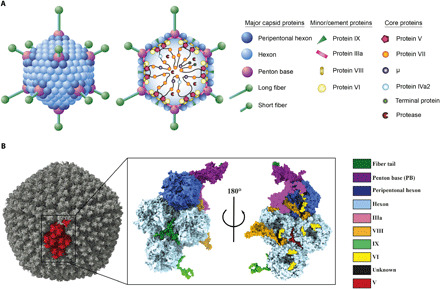
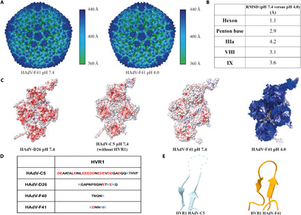
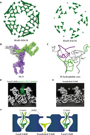
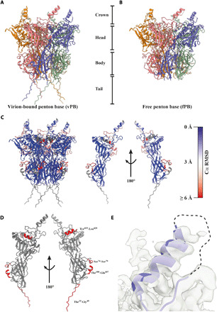
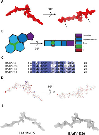
Human adenovirus (HAdV) types F40 and F41 are a prominent cause of diarrhea and diarrhea-associated mortality in young children worldwide. These enteric HAdVs differ notably in tissue tropism and pathogenicity from respiratory and ocular adenoviruses, but the structural basis for this divergence has been unknown. Here, we present the first structure of an enteric HAdV-HAdV-F41-determined by cryo-electron microscopy to a resolution of 3.8 Å. The structure reveals extensive alterations to the virion exterior as compared to nonenteric HAdVs, including a unique arrangement of capsid protein IX. The structure also provides new insights into conserved aspects of HAdV architecture such as a proposed location of core protein V, which links the viral DNA to the capsid, and assembly-induced conformational changes in the penton base protein. Our findings provide the structural basis for adaptation of enteric HAdVs to a fundamentally different tissue tropism.


Figures

Fig. 1
Fig. 1. The overall structure of HAdV-F41.
(A) Schematic representation of the capsid and core structure of HAdV-F41. (B) Surface representation of the HAdV-F41 electron density with one asymmetric unit (ASU) highlighted in red (left) and a surface representation of the ASU of the HAdV-F41 atomic model viewed from the virion exterior (middle) and interior (right).
Fig. 2
Fig. 2. Structure and surface charge distribution of HAdV-F41 at pH 7.4 and pH 4.0.
(A) Surface representation of the HAdV-F41 electron density at pH 7.4 and pH 4.0, colored by distance from the virion center. (B) pH-dependent structural changes in selected HAdV-F41 capsid proteins as measured by RMSD at Cα level. (C) Surface charge distribution for the atomic models of HAdV-C5 (PDB: 6CGV), HAdV-D26 (PDB: 5TX1), and HAdV-F41 at pH 7.4 as well as HAdV-F41 at pH 4.0. Red represents a local net negative charge, blue represents positive, and white represents uncharged. (D) Comparison of the HVR1 sequences between HAdV-C5, HAdV-D26, HAdV-F40, and HAdV-F41. (E) Cartoon representation of the HVR1-containing loop for HAdV-C5 and HAdV-F41. The unbuilt loop for HAdV-C5 is shown as a dashed line.
Fig. 3
Fig. 3. The triskelion-forming protein IX assembles in a unique way in HAdV-F41.
(A) Surface representation of IX assembly in HAdV-D26 and HAdV-F41 including all ordered protein density. (B) Graphical representation of HAdV-F41 IX-N triskelion assembly. The three IX chain atoms (green, gray, and purple) are shown in stick representation covered by a semitransparent surface. (C) Close-up view of the hydrophobic core at the center of the IX triskelion assembly, formed by residues Phe12, Phe17, and Tyr20 of each IX chain. (D) Computational slice through the electron density of HAdV-F41 at pH 7.4, at the position of IX at the local threefold axis. Electron density is white except the proposed density of IX-C, which is highlighted in green. (E) As (D), but at the icosahedral threefold axis. (F) Schematic representation of one possible arrangement of IX-C.
Fig. 4
Fig. 4. The HAdV-F41 PB undergoes assembly-induced conformational changes.
(A) Cartoon representation of the virion-bound PB (vPB), which can be divided into a crown, head, body, and tail. (B) Cartoon representation of the free PB (fPB). (C) Cartoon representation of the vPB and a single vPB monomer chain, each colored by Cα RMSD indicating the local degree of difference in Cα positioning between the vPB and fPB structures. (D) Cartoon representation of a single vPB monomer chain (gray). Missing residues in the fPB structure are highlighted in red. (E) Cartoon representation of the helix and disordered loop region containing the integrin-binding IGDD motif (dashed line) located in the crown. The electron density is shown as a transparent surface.
Fig. 5
Fig. 5. Location of DNA binding protein V at the interface of the three nonperipentonal hexon subunits.
(A) Surface representation of V electron density. Arrows indicate the positions fixed during bioinformatics analysis. (B) Schematic representation of V and its location in the HAdV-F41 ASU. (C) Alignment of the identified V amino acid sequence from HAdV-C5, HAdV-D26, HAdV-F40, and HAdV-F41. Coloring represents percent sequence identity, with dark blue illustrating 100% homology. (D) Graphical representation of the modeled HAdV-F41 V peptide, shown in maroon, and stick representation covered by the corresponding electron density, shown as transparent surface. (E) Surface representation of the HAdV-C5 [EMD-7034 (20)] and HAdV-D26 [EMD-8471 (21)] electron densities located at the same position in their respective ASUs.

References

    1. W. S. M. Wold, M. G. Ison, Adenoviruses, in Fields Virology, D. M. Knipe, P. M. Howley, Eds. (Lippincott Williams & Wilkins, 2016), vol. 2, chap. 56, pp. 1732–1767.
    1. HAdV Working Group, (2019); http://hadvwg.gmu.edu/.
    1. Garofalo M., Staniszewska M., Salmaso S., Caliceti P., Pancer K. W., Wieczorek M., Kuryk L., Prospects of replication-deficient adenovirus based vaccine development against SARS-CoV-2. Vaccines 8, 293 (2020). - PMC - PubMed
    1. Guo X., Deng Y., Chen H., Lan J., Wang W., Zou X., Hung T., Lu Z., Tan W., Systemic and mucosal immunity in mice elicited by a single immunization with human adenovirus type 5 or 41 vector-based vaccines carrying the spike protein of Middle East respiratory syndrome coronavirus. Immunology 145, 476–484 (2015). - PMC - PubMed
    1. Collaborators G. B. D. D. D., Estimates of the global, regional, and national morbidity, mortality, and aetiologies of diarrhoea in 195 countries: A systematic analysis for the Global Burden of Disease Study 2016. Lancet Infect. Dis. 18, 1211–1228 (2018). - PMC - PubMed

Publication types

LinkOut - more resources