. 2021 Feb 1;17(2):e1009262.
doi: 10.1371/journal.ppat.1009262. eCollection 2021 Feb.

Capture and visualization of live Mycobacterium tuberculosis bacilli from tuberculosis patient bioaerosols

Affiliations

Capture and visualization of live Mycobacterium tuberculosis bacilli from tuberculosis patient bioaerosols

Ryan Dinkele et al. PLoS Pathog. .

Abstract

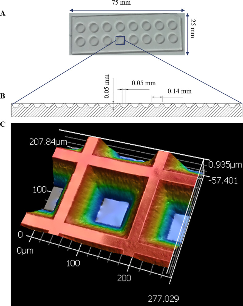
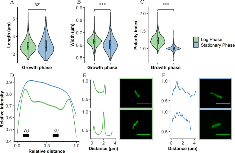
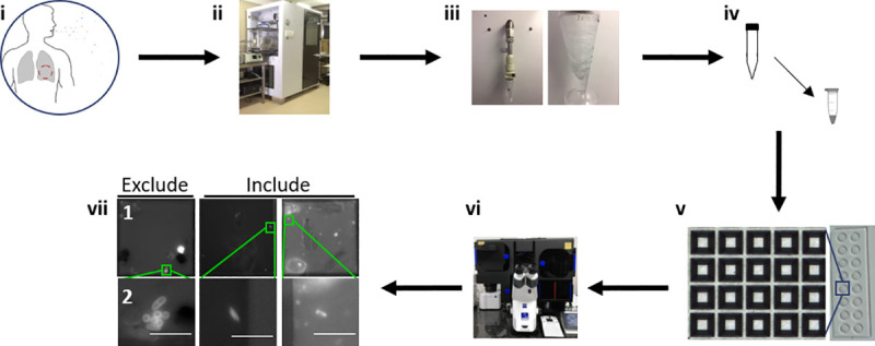
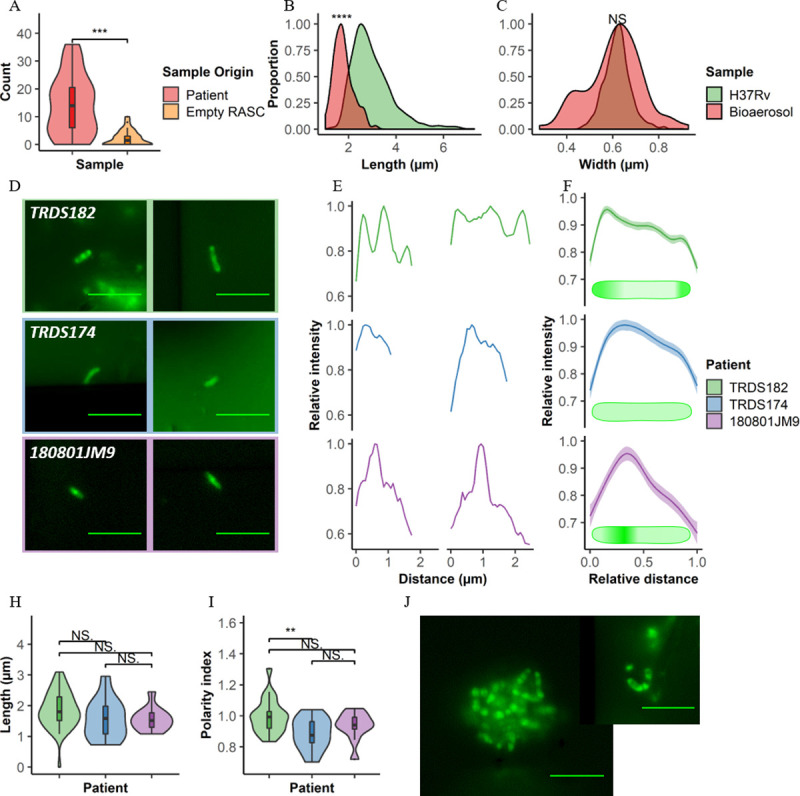
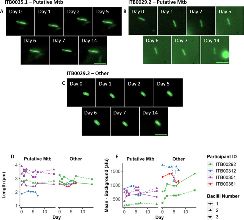
Interrupting transmission is an attractive anti-tuberculosis (TB) strategy but it remains underexplored owing to our poor understanding of the events surrounding transfer of Mycobacterium tuberculosis (Mtb) between hosts. Determining when live, infectious Mtb bacilli are released and by whom has proven especially challenging. Consequently, transmission chains are inferred only retrospectively, when new cases are diagnosed. This process, which relies on molecular analyses of Mtb isolates for epidemiological fingerprinting, is confounded by the prolonged infectious period of TB and the potential for transmission from transient exposures. We developed a Respiratory Aerosol Sampling Chamber (RASC) equipped with high-efficiency filtration and sampling technologies for liquid-capture of all particulate matter (including Mtb) released during respiration and non-induced cough. Combining the mycobacterial cell wall probe, DMN-trehalose, with fluorescence microscopy of RASC-captured bioaerosols, we detected and quantified putative live Mtb bacilli in bioaerosol samples arrayed in nanowell devices. The RASC enabled non-invasive capture and isolation of viable Mtb from bioaerosol within 24 hours of collection. A median 14 live Mtb bacilli (range 0-36) were isolated in single-cell format from 90% of confirmed TB patients following 60 minutes bioaerosol sampling. This represented a significant increase over previous estimates of transmission potential, implying that many more organisms might be released daily than commonly assumed. Moreover, variations in DMN-trehalose incorporation profiles suggested metabolic heterogeneity in aerosolized Mtb. Finally, preliminary analyses indicated the capacity for serial image capture and analysis of nanowell-arrayed bacilli for periods extending into weeks. These observations support the application of this technology to longstanding questions in TB transmission including the propensity for asymptomatic transmission, the impact of TB treatment on Mtb bioaerosol release, and the physiological state of aerosolized bacilli.


Conflict of interest statement

The authors have declared that no competing interests exist.

Figures

Fig 1
Fig 1. Design and fabrication of nanowell-arrayed microscope slides for the compartmentalization and visualization of TB bioaerosols.
(A) Photograph and (B) schematic of a nanowell-arrayed microscope slide. (C) 3D scan of a 207.840 x 277.029 μm section of the slide. Each device (25 mm x 75 mm) consists of two rows of eight round microwells machined from cast acrylic. The microwells are 6 mm in diameter and 2 mm deep. The nanowell film, which is bonded to the superstructure with UV-curing adhesive, is made from embossed COC film. The nanowells have side-wall angles of 35° and are 50 μm deep. The distance through the bottom of each well to the back of the film is ~170 μm, equivalent to a number 1.5 coverslip.
Fig 2
Fig 2. Differentiation of growth states in Mtb using DMN-trehalose and cell morphology.
Comparisons between log (green) and stationary (blue) phase Mtb according to (A) cell length, (B) width, and (C) polarity index. (D) Average DMN-trehalose profile for log and stationary phase bacilli, with single-cell examples in both (E) log and (F) stationary phase. Polarity index for each cell was calculated as the median fluorescence intensity at region (1) divided by the median fluorescence intensity at region (2) in panel (D). Wilcoxon signed-rank test performed, p < 0.001 = ***, NS = not significant.
Fig 3
Fig 3. Workflow from participant recruitment to image analysis.
(i) Recruitment of TB GeneXpert+ patients. (ii) One hour of bioaerosol production during tidal breathing and non-induced cough within the Respiratory Aerosol Sampling Chamber (RASC). (iii) Liquid capture of patient bioaerosol via Bertin Coriolis μ Biological Air Sampler. (iv) Bioaerosol concentration and staining with 100 μM DMN-trehalose during overnight (~16 hours) incubation at 37 oC. (v) Sample arraying within the nanowell device. (vi) Manual sample scanning and bacilli enumeration. (vii) Nanowell imaging (row 2 represents a zoomed in section from row 1). Columns represent 3 different patients. Bacilli not matching inclusion criteria are excluded from subsequent analysis. Scale bar, 5 μm.
Fig 4
Fig 4. Detection and characterization of putative Mtb within bioaerosols of confirmed TB patients.
(A) Plot comparing the number of putative Mtb detected within TB+ participants (red, n = 31) and empty RASC controls (orange, n = 27). Comparing distributions of (B) cell lengths and (C) cell widths in putative Mtb bacilli detected within bioaerosols of TB patients (red) to Mtb H37Rv cultured within the lab (green). Representative (D) images and (E) plots of the three distinct, exemplar cytological profiles from three patients in which putative Mtb were detected. (F) Average plots and idealised drawings indicating the different staining patterns of all bacilli detected in these three patients. (H) Polarity index and (I) length of the bacilli detected within these patients. (J) Representative images of clumps of putative Mtb detected within bioaerosol samples (TRDS182). Scale bar = 5 μm. Wilcoxon Rank-Sum test performed, p < 0.01 = **, < 0·001 = ***, p < 0,0001 = ****, NS = not significant.
Fig 5
Fig 5. Serial imaging of organisms captured directly from patient bioaerosol for up to two weeks.
Single bacilli identified from 4 separate patients were serially imaged daily for the first 7 days (except weekends) and weekly thereafter. Up to three bacilli were tracked per patient (bacilli number–represented by shape and dashed lines) in four patients (represented by colour) and identified as either “Putative Mtb” or “Other”. (A—C) Representative bacilli from two separate patients imaged on days 0, 1, 2, 5, 6, 7, and 14. (A) and (B) represent putative Mtb, whereas (C) represents other organisms with a low probability of being Mtb based on the applied inclusion criteria. Summary of bacterial changes in (D) length and (E) mean fluorescence intensity minus the average background intensity. Scale bar = 5μm.

References

    1. World Health Organization. Global Tuberculosis Report. 2019.
    1. Kaplan G. Tuberculosis control in crisis-causes and solutions. Prog Biophys Mol Biol. 2019; published online Oct 15. 10.1016/j.pbiomolbio.2019.10.001 - DOI - PubMed
    1. Sharma A, Hill A, Kurbatova E, van der Walt M, Kvasnovsky C, Tupasi T et al. Estimating the future burden of multidrug-resistant and extensively drug-resistant tuberculosis in India, the Philippines, Russia, and South Africa: a mathematical modelling study. Lancet Infect Dis. 2017; 17:707–715. 10.1016/S1473-3099(17)30247-5 - DOI - PMC - PubMed
    1. Koch A, Cox H. Preventing drug-resistant tuberculosis transmission. Lancet Infect Dis. 2019; published online Nov 26. 10.1016/S1473-3099(19)30613-9 - DOI - PubMed
    1. Glynn JR, Guerra-Assunção JA, Houben RMGJ, Sichali L, Mzembe T, Mwaungulu LK, et al. Whole genome sequencing shows a low proportion of tuberculosis disease is attributable to known close contacts in rural Malawi. PLoS One. 2015; 10: 1–12. - PMC - PubMed

Publication types