. 2021 Feb 1;16(2):e0246050.
doi: 10.1371/journal.pone.0246050. eCollection 2021.

Pseudomonas aeruginosa induces p38MAP kinase-dependent IL-6 and CXCL8 release from bronchial epithelial cells via a Syk kinase pathway

Affiliations

Pseudomonas aeruginosa induces p38MAP kinase-dependent IL-6 and CXCL8 release from bronchial epithelial cells via a Syk kinase pathway

Matthew S Coates et al. PLoS One. .

Abstract

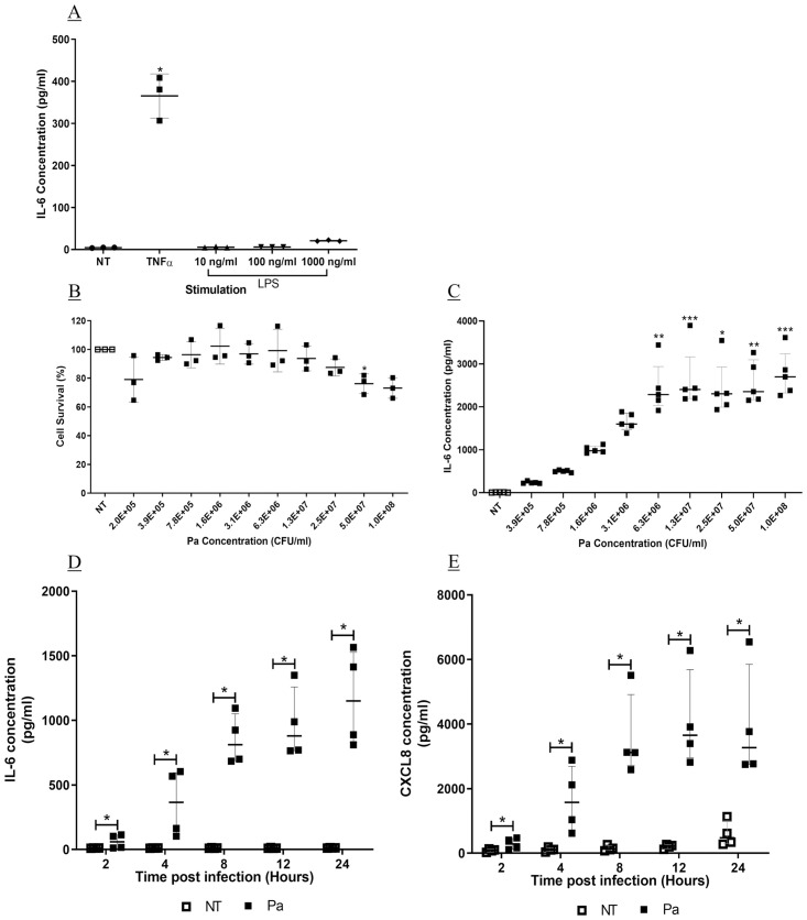
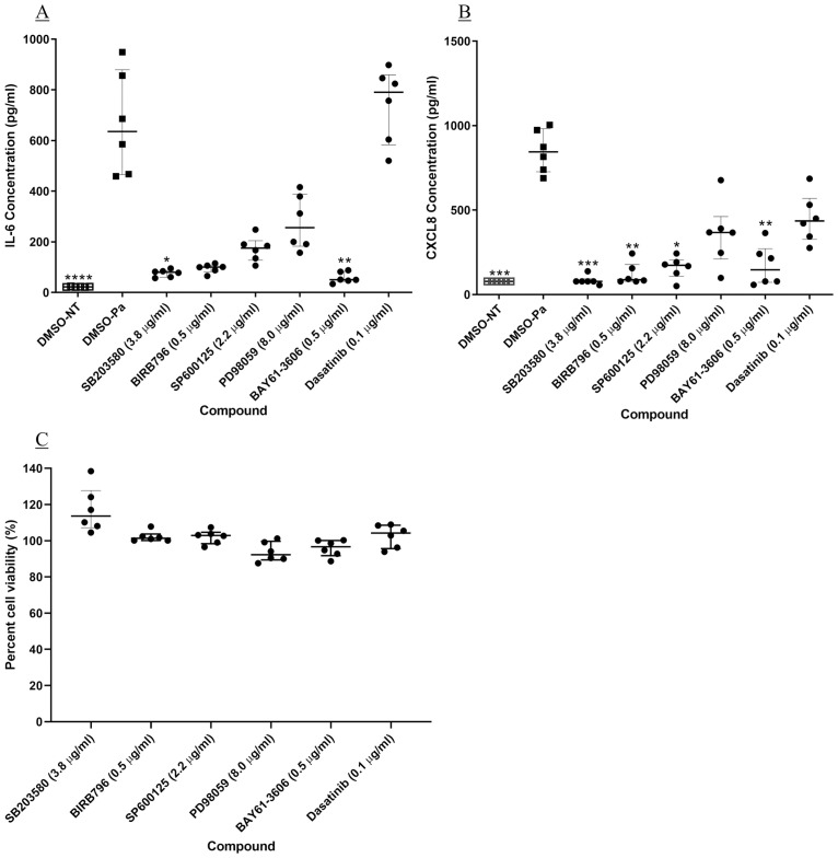
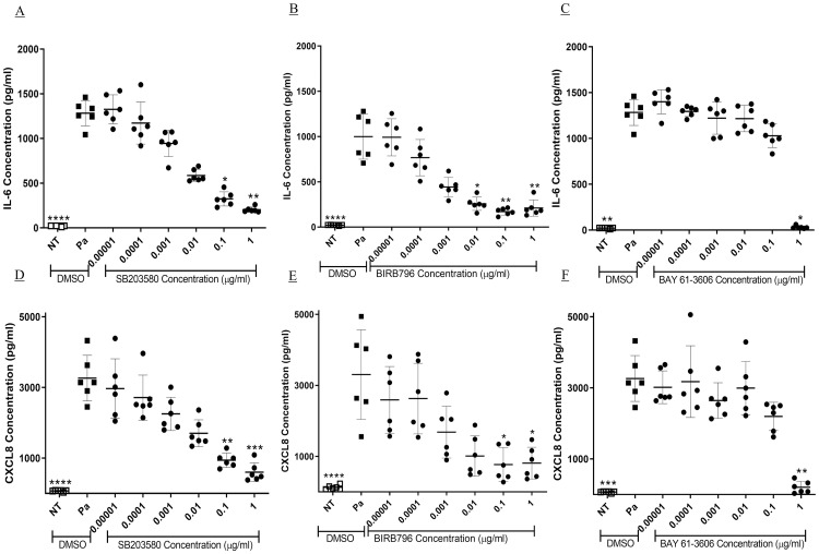
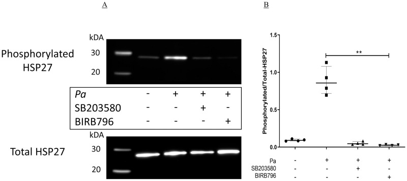
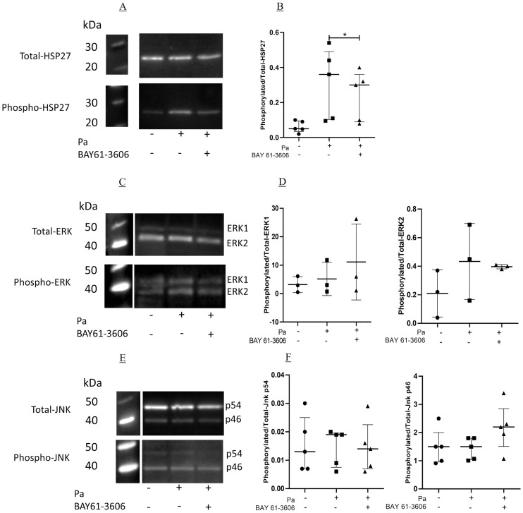
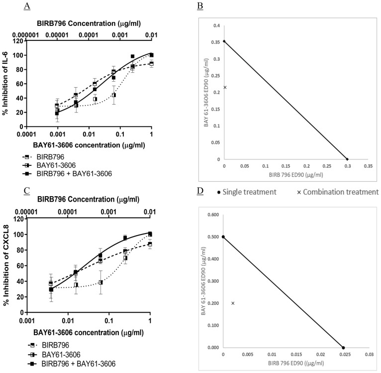
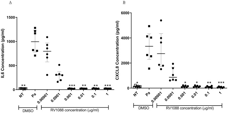
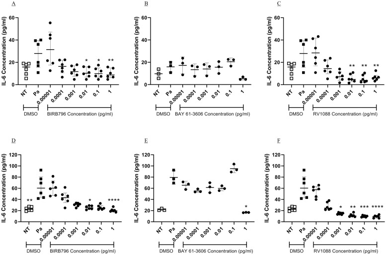
Pseudomonas aeruginosa (Pa) infection is a major cause of airway inflammation in immunocompromised and cystic fibrosis (CF) patients. Mitogen-activated protein (MAP) and tyrosine kinases are integral to inflammatory responses and are therefore potential targets for novel anti-inflammatory therapies. We have determined the involvement of specific kinases in Pa-induced inflammation. The effects of kinase inhibitors against p38MAPK, MEK 1/2, JNK 1/2, Syk or c-Src, a combination of a p38MAPK with Syk inhibitor, or a novel narrow spectrum kinase inhibitor (NSKI), were evaluated against the release of the proinflammatory cytokine/chemokine, IL-6 and CXCL8 from BEAS-2B and CFBE41o- epithelial cells by Pa. Effects of a Syk inhibitor against phosphorylation of the MAPKs were also evaluated. IL-6 and CXCL8 release by Pa were significantly inhibited by p38MAPK and Syk inhibitors (p<0.05). Phosphorylation of HSP27, but not ERK or JNK, was significantly inhibited by Syk kinase inhibition. A combination of p38MAPK and Syk inhibitors showed synergy against IL-6 and CXCL8 induction and an NSKI completely inhibited IL-6 and CXCL8 at low concentrations. Pa-induced inflammation is dependent on p38MAPK primarily, and Syk partially, which is upstream of p38MAPK. The NSKI suggests that inhibiting specific combinations of kinases is a potent potential therapy for Pa-induced inflammation.


Conflict of interest statement

I have read the journal’s policy and the authors of this manuscript have the following competing interests: Previously Matthew S. Coates was employed by Respivert Ltd. Garth W. Rapeport and Kazuhiro Ito were co-founders and employees of Respivert Ltd. During the period of the research G. W. Rapeport and K. Ito were co-founders and employees of Pulmocide Ltd. This does not alter our adherence to PLOS ONE policies on sharing data and materials.

Figures

Fig 1
Fig 1. Stimulation of IL-6 and CXCL8 from BEAS-2B cells.
BEAS-2B cells were stimulated with (A) TNFα at 10 ng/ml or LPS at 10, 100 or 100 ng/ml for four hours. Cell free supernatant was collected, and IL-6 measured by sandwich ELISA (n = 3, mean ± SD). (B) BEAS-2B cells were infected with Pa at varying concentrations for one hour, followed by addition of gentamicin at 100 μg/ml and a further 24 hours incubation. Cell viability was measured by MTT assay (n = 3, mean ± SD). (C) Cell free supernatant from cells infected with Pa for 24 hours was analysed for IL-6 by sandwich ELISA (n = 5, median with IQR). BEAS-2B cells were infected with Pa at 2.5 x 10⁷ CFU/ml for one hour, followed by addition of gentamicin at 100 μg/ml. Supernatant was collected at 2, 4, 8, 12 or 24 hours later. (D) IL-6 and (E) CXCL8 were measured by sandwich ELISA (n = 4, median with IQR). (A-C) Stimulants were compared to NT using Friedman test with Dunn’s correction. (D-E) At each time point, Pa treatments were compared with NT using the Mann-Whitney U test (* = p<0.05** = p<0.01, *** = p<0.001).
Fig 2
Fig 2. Head to head comparison of kinase inhibitors against Pa-induced IL-6 and CXCL8 release, and cell viability.
BEAS-2B cells underwent a two-hour pre-treatment with kinase inhibitors at a single concentration known to completely inhibit the target kinase enzyme activity. The cells were stimulated with Pa at 2.5 x 10⁷ CFU/ml for one hour followed by addition of gentamicin at 100 μg/ml and a further four-hour incubation. The concentration of (A) IL-6 and (B) CXCL8 in the cell-free supernatant was measured by sandwich ELISA. (C) BEAS-2B cell viability was measured after compound treatment and Pa infection, using an MTT assay; cell viability was compared to cells treated with DMSO and Pa. Friedman test with Dunn’s multiple comparisons was used to compare each kinase inhibitor with vehicle treatment. n = 6, showing median with IQR (* = p<0.05, ** = p<0.01 and *** = p<0.001). Compound concentrations: SB203580–3.8 μg/ml; BIRB796–0.5 μg/ml; SP600125–2.2 μg/ml; PD98059–8.0 μg/ml; BAY 61–3606–0.5 μg/ml and Dasatinib– 0.1 μg/ml.
Fig 3
Fig 3. Concentration dependent inhibition of Pa-induced IL-6 and CXCL8 release by selected kinase inhibitors.
BEAS-2B cells underwent a two-hour pre-treatment with 10-fold dilutions of (A and D) SB203580, (B and E) BIRB796 and (C and F) BAY 61–3606. The cells were stimulated with Pa at 2.5 x 10⁷ CFU/ml for one hour followed by addition of gentamicin at 100 μg/ml and a further four-hour incubation. The concentration of (A, B and C) IL-6 and (D, E and F) CXCL8 in the cell free supernatant was measured by sandwich ELISA. Friedman test with Dunn’s multiple comparisons was used to compare each kinase inhibitor with vehicle treatment. n = 6, showing median with IQR (* = p<0.05, ** = p<0.01 and *** = p<0.001).
Fig 4
Fig 4. Inhibition of Pa-induced phosphorylation of HSP27 by p38MAPK inhibitors.
BEAS-2B cells in six well plates were pre-incubated with either DMSO (NT), SB203580 or BIRB796 at 1 μg/ml for two hours at 37°C, 5% CO2. The cells were then stimulated with Pa at 2.5 x 10⁷ CFU/ml for two hours at 37°C, 5% CO2, after which whole cell protein was collected and levels of phosphorylated- and total-HSP27 were measured by Western blot. (A) Representative image of the four experiments. (B) Band intensity was determined using Syngene Gene Tools software and the levels of phosphorylated-HSP27 were corrected to the levels of total-HSP27. A Friedman test with Dunn’s multiple comparison was used to compare Pa treatment alone with all other conditions. n = 4 showing median with IQR (** = p≤0.01).
Fig 5
Fig 5. Inhibition of MAP kinase activity by BAY 61–3606.
BEAS-2B cells in six well plates were pre-incubated with either DMSO, or BAY 61–3606 at 1 μg/ml for two hours at 37°C, 5% CO2. The cells were then incubated with Pa at 2.5 x 10⁷ CFU/ml for two hours at 37°C, after which whole cell protein was collected. Western blots were carried out to measure the levels of total and phosphorylated (A) HSP27, (C) ERK and (E) JNK. (B, D and F) Band intensities were calculated using Syngene Gene Tools and phosphorylated protein levels were corrected to total protein. BAY 61–3606 treatment was compared to DMSO using a one-tailed Wilcoxon sign ranked test. (B) and (F), n = 5, median with IQR, (D) n = 3, mean ± SD. Images are representative of the repeats conducted (* = p<0.05).
Fig 6
Fig 6. Synergy of p38MAPK and Syk inhibition against Pa-induced IL-6 and CXCL8 release.
BEAS-2B cells underwent a two-hour pre-treatment with four-fold dilutions of either BIRB796 or BAY 61–3606, or a combination of the two. The cells were stimulated with Pa at 2.5 x 10⁷ CFU/ml for one hour followed by addition of gentamicin at 100 μg/ml and a further four-hour incubation. The concentration of (A) IL-6 and (C) CXCL8 in the cell free supernatant were measured by sandwich ELISA, n = 3, showing mean ± SD. Synergy was calculated using the Chou-Talalay method using CompuSyn software [64]. Isobolograms of the ED90 concentrations for the compound concentrations against (B) IL-6 and (D) CXCL8 were drawn using Microsoft Excel. The ED90 of each compound applied alone is plotted on the x and y axes and joined with a line. The ED90 values of each compound when used in combination are plotted as an X. An X on the line would indicate an additive effect; X on the left of the line would indicate synergy (as in these experiments), and to the right would indicate compound antagonism.
Fig 7
Fig 7. Inhibition of IL-6 and CXCL8 by a novel narrow spectrum kinase inhibitor.
BEAS-2B cells underwent a two-hour pre-treatment with 10-fold dilutions of RV1088. The cells were stimulated with Pa at 2.5 x 10⁷ CFU/ml for one hour followed by addition of gentamicin at 100 μg/ml and a further four-hour incubation. The concentration of (A) IL-6 and (B) CXCL8 in the cell free supernatant was measured by sandwich ELISA. Friedman test with Dunn’s multiple comparisons was used to compare each kinase inhibitor with vehicle treatment. n = 6, showing median with IQR (* = p<0.05, ** = p<0.01 and *** = p<0.001).
Fig 8
Fig 8. Inhibition of Pa-induced IL-6 from bronchial epithelial cells expressing Phe508del- and WT-CFTR.
CFBE41o- cells expressing either Phe508del (A-C) or WTCFTR (D-F) underwent a two-hour pre-treatment with 10-fold dilutions of BIRB796 (A and D), BAY 61–3606 (B and E) and RV1088 (C and E). The cells were stimulated with Pa at 2.5 x 10⁷ CFU/ml for one hour followed by addition of gentamicin at 100 μg/ml and a further four-hour incubation. The concentration of IL-6 in the cell free supernatant was measured by sandwich ELISA. Friedman test with Dunn’s multiple comparisons was used to compare each kinase inhibitor with vehicle treatment. n = 6 for BIRB796 and RV1088, showing median with IQR, n = 3 for BAY 61–3606, showing mean and SD (* = p<0.05, ** = p<0.01 and *** = p<0.001, **** = p<0.0001).

Similar articles

Cited by

References

    1. Gellatly SL, Hancock REW. Pseudomonas aeruginosa: new insights into pathogenesis and host defenses. Pathog Dis. 2013;67: 159–173. 10.1111/2049-632X.12033 - DOI - PubMed
    1. Coburn B, Wang PW, Diaz Caballero J, Clark ST, Brahma V, Donaldson S, et al. Lung microbiota across age and disease stage in cystic fibrosis. Sci Rep. 2015;5: 10241 10.1038/srep10241 - DOI - PMC - PubMed
    1. Kosorok MR, Zeng L, West SE, Rock MJ, Splaingard ML, Laxova A, et al. Acceleration of lung disease in children with cystic fibrosis after Pseudomonas aeruginosa acquisition. Pediatr Pulmonol. 2001;32: 277–287. 10.1002/ppul.2009.abs - DOI - PubMed
    1. Pier GB. Pseudomonas aeruginosa lipopolysaccharide: a major virulence factor, initiator of inflammation and target for effective immunity. Int J Med Microbiol. 2007;297: 277–295. 10.1016/j.ijmm.2007.03.012 - DOI - PMC - PubMed
    1. Soong G, Reddy B, Sokol S, Adamo R, Prince A. TLR2 is mobilized into an apical lipid raft receptor complex to signal infection in airway epithelial cells. J Clin Invest. 2004;113: 1482–1489. 10.1172/JCI20773 - DOI - PMC - PubMed

Publication types