Review
. 2021 Aug 17;45(4):fuab004.
doi: 10.1093/femsre/fuab004.

Archaeal tyrosine recombinases

Affiliations
Review

Archaeal tyrosine recombinases

Catherine Badel et al. FEMS Microbiol Rev. .

Abstract

The integration of mobile genetic elements into their host chromosome influences the immediate fate of cellular organisms and gradually shapes their evolution. Site-specific recombinases catalyzing this integration have been extensively characterized both in bacteria and eukarya. More recently, a number of reports provided the in-depth characterization of archaeal tyrosine recombinases and highlighted new particular features not observed in the other two domains. In addition to being active in extreme environments, archaeal integrases catalyze reactions beyond site-specific recombination. Some of these integrases can catalyze low-sequence specificity recombination reactions with the same outcome as homologous recombination events generating deep rearrangements of their host genome. A large proportion of archaeal integrases are termed suicidal due to the presence of a specific recombination target within their own gene. The paradoxical maintenance of integrases that disrupt their gene upon integration implies novel mechanisms for their evolution. In this review, we assess the diversity of the archaeal tyrosine recombinases using a phylogenomic analysis based on an exhaustive similarity network. We outline the biochemical, ecological and evolutionary properties of these enzymes in the context of the families we identified and emphasize similarities and differences between archaeal recombinases and their bacterial and eukaryal counterparts.

Keywords: Archaea; genome evolution; horizontal transfer; mobile genetic element; site-specific recombination; tyrosine recombinase.


Figures

Figure 1.
Figure 1.
Campbell model for phage λ site-specific recombination. The model presented by Campbell (1963) suggested for the first time the breaking and rejoining of DNA sequences by integration enzymes in order to allow phage λ lysogenization in Escherichia coli.
Figure 2.
Figure 2.
Classes of archaeal integrases. She, Brugger and Chen (2002) proposed the ranking of archaeal integrases into two distinct types: Type I (A) and Type II (B). The blue and red squares correspond to the specific recombination sites.
Figure 3.
Figure 3.
Archaeal integrases sequence domains and conserved residues.(A) Suicidal integrases are either encoded by their intact gene or the regions corresponding to the N-terminal portion or Int(N), and C-terminal region or Int(C) are separated upon MGE integration. (B) The conserved catalytic residues are indicated for all characterized archaeal tyrosine integrases from Table 1. The domains of particular interest are indicated. Functional domains were dissected for IntSSV2 (Zhan, Zhou and Huang 2015). IntpTN3 presents additional loop that may be responsible for its unprecedented dual catalytic activity (Cossu et al. 2017). PaXerA and TaXerA structure was resolved and corresponds to two domains separated by a linker (Serre et al. ; Jo et al. 2016).
Figure 4.
Figure 4.
Classification of archaeal tyrosine recombinases. Alluvial diagram showing the integrase distribution across the different archaeal tyrosine recombinase superfamilies (left), families (center) and subfamilies (right), predicted based on the conservation identity threshold of 25%, 30% and 45%, respectively, using SiLiX (Miele, Penel and Duret 2011). In this diagram, blocks represent clusters of proteins and stream fields between the blocks represent changes in clustering attribution of these proteins to superfamily, family and subfamily over the selected identity threshold. Block height is proportional to the size of the protein cluster and the height of a stream field is proportional to the number of proteins contained within the blocks connected by the stream field. Our classification retained the 17 families containing >13 members. The graph was drawn using RawGraphs (https://app.rawgraphs.io) to map data dimensions onto visual variables. The raw data are available in Table S1 (Supporting Information).
Figure 5.
Figure 5.
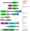
Pfam domain combinations in the 17 major tyrosine recombinase families. The most frequent combinations of Pfam domains for each major tyrosine recombinase are represented. No Pfam could be detected for the fam30-08 and STSV/SMV families even if both belong to the cd00397 superfamily. The result of the conserved domain search is available in Table S1 (Supporting Information).
Figure 6.
Figure 6.
Distribution of the 17 major tyrosine recombinase families among the archaeal diversity. Each tyrosine recombinase family corresponds to a dot. In order to visualize the distribution of the major tyrosine recombinase families on the archaeal diversity, we used data from AnnoTree (Mendler et al. 2019) with additional manual curation. The archaeal tree was obtained from the Genome Taxonomy Database (GTDB Release 03-RS86) (Parks et al. 2020), generated from 122 core proteins and exported using taxonomic orders as resolution level. The presence/absence profiles for each family were visualized using iTOL (Letunic and Bork 2019).
Figure 7.
Figure 7.
Similarity network of archaeal tyrosine recombinases. A similarity network performed with SiLiX (Miele, Penel and Duret 2011) and visualized with Igraph (https://igraph.org) was used to assign tyrosine recombinases from the superfamily Superfam25-02 to families and subfamilies. The similarities of all against all proteins of the dataset were assessed using BlastP (expect >0.001, with an identity threshold of 30% among 60% of the protein). Protein clustering was achieved by a random walk algorithm. Each circle corresponds to an individual protein colored according to the clustering.
Figure 8.
Figure 8.
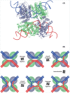
Tyrosine recombinase model and site-specific recombination reaction. (A) Structural model of the Cre-loxA pre-synapse complex. The tetrameric conformation of the Cre-loxA pre-synapse is clearly apparent with its active cleaving subunits in green color. The DNA components of the recombination complex are shown in blue and red backbone form. The tridimensional model is referenced as PDB 1NZB (Guo, Gopaul and van Duyne 1997). (B)Site-specific recombination model. Schematic representation of the consecutive reactions leading to the formation of recombinant DNA molecules by tyrosine recombinases. The color code is consistent with that of Panel A. The active tyrosine residues are shown in purple color.
Figure 9.
Figure 9.
The different outcomes of site-specific recombination. The red and blue rectangles correspond to the specific recombination sites. With circular DNA molecules as substrates, the recombination outcome can be (A) integration and excision or (B)inversion, depending on the relative position of the two specific sites. (C)Recombination between two linear DNA molecules results in two chimeric linear DNA molecules.
Figure 10.
Figure 10.
Assays to detect integrase activity in vitro and in vivo.(A) Different substrates harboring specific sites (light blue arrows) are incubated with the integrases in vitro and the products were monitored (Cortez et al. ; Zhan, Zhou and Huang ; Cossu et al. ; Jo et al. ; Badel et al. 2020). (B) The half-site strand transfer assay was first implemented in vitro by Serre et al. (2013). It allows the verification of the strand cleavage site (in red). The incubation of the integrase with half of the specific site results in a covalent DNA–protein complex that can be detected (left). The incubation of the integrase with two separated halves of the specific site results in the reconstruction of the entire specific site only if the substrate halves were designed accordingly to the cleavage site (right). (C) A non-replicative plasmid harboring the specific site and a selectable marker is introduced in a suitable host cell. Upon selection, only the cells where the integrase catalyzes plasmid integration can grow (Wang et al. 2018). (D) A replicative plasmid harboring two specific sites and a split selectable marker is introduced into the appropriate cell. The selectable marker is reconstituted only if the integrase catalyzes recombination between the two specific sites. The cell can then grow upon selection (Wang et al. 2018). (E) Different arrangements of the specific site result from integration or excision. They can be detected by polymerase chain reaction with different pairs of four primers (red and green arrows) (Li et al. ; Cossu et al. ; Wang et al. 2018).
Figure 11.
Figure 11.
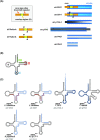
Archaeal tyrosine recombinase recombination sites.(A) Recombination sites are sketched for the characterized archaeal tyrosine recombinases. The orange and yellow boxes correspond to core-type sites as defined for bacteriophage λ (Landy 2015). The blue box corresponds to the att site. The black sequences are necessary and sufficient for recombination in vitro (Cossu et al. ; Badel et al. 2020). The black arrows indicate the cleavage site when experimentally determined (Serre et al. , ; Jo et al. 2017). (B) General organization of a tRNA indicating the location of the T, A and D loops and the three preferred integration locations for bacterial integrases (I, II and III) (Williams 2002). (C) Att sites often correspond to tRNA sequences. The leaf-like structure of the targeted tRNA is indicated with att site nucleotides circled in blue.
Figure 12.
Figure 12.
Wide tRNA genes targeting by archaeal tyrosine recombinases. All tRNA anticodon combinations are listed along with their corresponding amino acids. Anticodons that are not found in archaeal tRNAs are indicated in light gray. An array of symbols indicates the utilization of a tRNA gene with the specified anticodon as attB site for Thermococcales (Li et al. ; Cossu et al. ; Badel et al. 2019, 2020), Halobacteriales (Krupovic, Forterre and Bamford ; Liu et al. 2015), Archaeoglobales (Badel et al. 2020), Methanococcales (Krupovic, Forterre and Bamford ; Badel et al. 2019), Methanosarcinales (Badel et al. 2020), Sulfolobales (She, Brugger and Chen ; Wang et al. ; Peng ; Redder et al. 2009) and Thaumarchaeota (Krupovic et al. 2019). Anticodons corresponding to untargeted tRNA genes are highlighted in bold green.
Figure 13.
Figure 13.
Suicidal integrase recombination. The integration by site-specific recombination (SSR) of multiple related MGE at different chromosomal locations and in inverted orientation (A) gives rise to homologous recombination (HR) between conserved MGE sequences (B, C). Such a recombination has been observed in T. kodakarensis (Gehring et al. 2017). Recombinant integrated MGEs encoding hybrid integrases can then excise if a compatible integrase is provided by a superinfecting MGE (D).

Similar articles

Cited by

References

    1. Abremski K, Gottesman S. Purification of the bacteriophage lambda Xis gene product required for lambda excisive recombination. J Biol Chem. 1982;257:9658–62. - PubMed
    1. Abremski K, Wierzbicki A, Frommer Bet al. . Bacteriophage P1 Cre-loxP site-specific recombination. Site-specific DNA topoisomerase activity of the Cre recombination protein. J Biol Chem. 1986;261:391–6. - PubMed
    1. Abremski KE, Hoess RH. Evidence for a second conserved arginine residue in the integrase family of recombination proteins. Protein Eng. 1992;5:87–91. - PubMed
    1. Argos P, Landy A, Abremski Ket al. . The integrase family of site-specific recombinases: regional similarities and global diversity. EMBO J. 1986;5:433–40. - PMC - PubMed
    1. Arinkin V, Smyshlyaev G, Barabas O. Jump ahead with a twist: DNA acrobatics drive transposition forward. Curr Opin Struct Biol. 2019;59:168–77. - PMC - PubMed

Publication types