. 2021 Feb 26;49(4):2317-2332.
doi: 10.1093/nar/gkab029.

Insight into formation propensity of pseudocircular DNA G-hairpins

Affiliations

Insight into formation propensity of pseudocircular DNA G-hairpins

Martina Lenarčič Živković et al. Nucleic Acids Res. .

Abstract

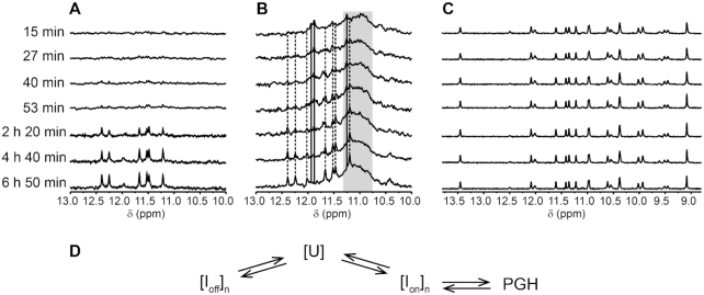
We recently showed that Saccharomyces cerevisiae telomeric DNA can fold into an unprecedented pseudocircular G-hairpin (PGH) structure. However, the formation of PGHs in the context of extended sequences, which is a prerequisite for their function in vivo and their applications in biotechnology, has not been elucidated. Here, we show that despite its 'circular' nature, PGHs tolerate single-stranded (ss) protrusions. High-resolution NMR structure of a novel member of PGH family reveals the atomistic details on a junction between ssDNA and PGH unit. Identification of new sequences capable of folding into one of the two forms of PGH helped in defining minimal sequence requirements for their formation. Our time-resolved NMR data indicate a possibility that PGHs fold via a complex kinetic partitioning mechanism and suggests the existence of K+ ion-dependent PGH folding intermediates. The data not only provide an explanation of cation-type-dependent formation of PGHs, but also explain the unusually large hysteresis between PGH melting and annealing noted in our previous study. Our findings have important implications for DNA biology and nanotechnology. Overrepresentation of sequences able to form PGHs in the evolutionary-conserved regions of the human genome implies their functionally important biological role(s).


Figures

Figure 1.
Figure 1.
Schematic representations of (A) G-hairpin, a proposed G-quadruplex folding intermediate, (B) a model of a G-hairpin imposed by DNA origami framework, and (C) a pseudocircular G-hairpin (PGH) adopted by SC11 sequence (PDB ID: 5M1W). Characteristic structural features of PGH fold include a chain reversal, responsible for mixed parallel/antiparallel backbone progression (red ellipse) and 5′-to-3′ stacking of terminal residues (dark green ellipse). G:G pairs are represented with dark grey, while loop regions are light grey.
Figure 2.
Figure 2.
Imino regions of 1D 1H NMR spectra of SC11 and its extended sequences.
Figure 3.
Figure 3.
(A) Imino region of the 1D 1H NMR spectrum of SC14 with the assigned imino proton resonances (top). Aromatic-imino (middle) and imino-imino (bottom) regions of the 2D NOESY spectrum (τm = 150 ms). The assignments of the aromatic and imino protons of the G:G core-forming residues are indicated on the right side of the NOESY spectrum in gray and black, respectively. The H8-H1 NOEs characteristic of N1-carbonyl, N7-amino base pair arrangements are marked with gray circles. (B) Schematic of N1-carbonyl symmetric (left) and N1-carbonyl, N7-amino (middle and right) hydrogen bond arrangements in a Hoogsteen–Watson–Crick (H-WC)-type and Watson–Crick–Hoogsteen (WC-H)-type arrangements, respectively. The aromatic-imino and imino-imino NOEs characteristic of the individual base pairing arrangement are indicated by gray and black arrows, respectively. (C) Aromatic-anomeric (top) and aromatic-aromatic (bottom) regions of the NOESY spectrum (τm = 150 ms) of SC14 in 2H2O. The sequential walk is indicated by solid lines. The missing cross-peaks are designated with asterisks, while respective correlations are indicated by dashed lines. Intraresidual H1′–H8 NOE cross-peaks belonging to guanines in anti and syn glycosidic conformations are depicted in dark and light blue, respectively. The cross-peaks indicative of a chain reversal, the formation of a new discontinuous G-tract and a flexible 3′-tail are shown in green, dark red and orange, respectively. (D) Schematic presentation of PGH topology adopted by SC14. The anti and syn G:G base pair-forming guanines are shown in dark and light blue, respectively. The loop residues and residues forming the flexible 3′-tail are colored in gray.
Figure 4.
Figure 4.
(A) Stereo-view of superposition of the 10 lowest-energy solution structures of SC14 PGH (PDB ID: 6R8E). (B) Arrangements of the G5:G1, G7:G−II and G6:G9 base pairs in the lowest-energy solution-state NMR structure. The hydrogen bonds are marked with dashed lines. (C) A junction between the pseudocircular element and the single stranded 3′-end protrusion in the SC14 PGH structure. The junction is stabilized by stacking of G9 and T10 and by hydrogen bonds between the N3 and O5′ atoms of G−II and the amino proton of G9. The guanines in anti and syn glycosidic conformations are shown in dark and light blue, respectively. The loop residues and residues G11 and T+I, which form the flexible 3′-tail are colored grey. O4′ atoms are colored red.
Figure 5.
Figure 5.
(A) Schematic representations of G:G cores in form I and form II PGHs. Note that both forms share the same distribution of syn (s) and anti (a) glycosidic conformations of guanines of the G:G core. The 5′-G involved in a core G:G base pair is marked with a red filled circle. Note: For the sake of simplicity, the chain-reversal element is not displayed. (B) Imino regions of 1D 1H NMR spectra of SC11-based constructs bearing 4′-alkoxy modification at indicated positions. (C) Imino region of 1D 1H NMR (left) and CD spectrum (right) of RNA counterpart of SC11, 5′-r(GUGUGGGUGUG)-3′.
Figure 6.
Figure 6.
Imino regions of 1D 1H NMR spectra of SC14 and its truncated variants, namely ΔT+I, ΔG11T+I and ΔT10G11T+I.
Figure 7.
Figure 7.
Schematic representation of the sequence requirements for form I (left) and form II (right) PGH formation in relation to their position in the PGH structure (A). Residues labeled with red and green circles correspond to the nucleotides critical and non-critical to PGH formation, respectively. (B) Sequence logos of minimal sequences capable to fold into form I (left) and form II (right) PGHs. For details on sequential logos determination, see Materials and Methods.
Figure 8.
Figure 8.
Imino regions of 1D 1H NMR spectra of a minimal PGH forming motifs (form I left, form II right) with their surroundings in human genome sorted by occurrence frequency.
Figure 9.
Figure 9.
(A) Imino regions of 1D 1H NMR spectra of SC11 acquired at 10°C. Samples were folded in the presence of different ratios (indicated) of NaCl and KCl. (B) Imino regions of 1D 1H NMR spectra of SC11 (top) and SC14 (bottom) acquired at 10°C in a buffer emulating the ion composition of the intracellular space (25 mM KPOi, pH 7, 110 mM KCl, 40 mM NaCl, 1 mM MgCl2 and 130 nM CaCl2).
Figure 10.
Figure 10.
(A and B) Imino regions of 1D 1H NMR spectra of SC11 acquired at 20°C and 1°C as a function of the time after annealing and quenching on ice in potassium phosphate based buffer (10 mM KPOi, pH 7, 100 mM KCl), respectively. Solid and dashed lines are indicative of signal positions of putative K+ ion-dependent intermediate (Ion) and those corresponding to PGH, respectively. Gray rectangle marks the spectral region with overlapped non-resolved signals presumed to correspond to off-folding pathway G-quadruplex-like intermediate(s) (Ioff). (C) Imino regions of 1D 1H NMR spectra of SC11 acquired at 1°C as a function of the time after annealing and quenching on ice in the sodium phosphate based buffer (10 mM NaPOi, pH 7, 100 mM NaCl). (D) Schematic/Simplistic representation of the proposed mechanism of PGH folding. U, Ion and Ioff stand for unfolded ensemble, K+ ion-dependent on-folding pathway intermediate(s), and off-folding pathway G-quadruplex like intermediate(s)/product(s), respectively. n ≥ 1. Note: The structure of species formed under Na+-based conditions has not been investigated in detail as it goes beyond the scope of the present study.

Similar articles

Cited by

References

    1. Mashimo T., Yagi H., Sannohe Y., Rajendran A., Sugiyama H.. Folding pathways of human telomeric type-1 and type-2 G-Quadruplex structures. J. Am. Chem. Soc. 2010; 132:14910–14918. - PubMed
    1. Gray R.D., Trent J.O., Chaires J.B.. Folding and unfolding pathways of the human telomeric G-quadruplex. J. Mol. Biol. 2014; 426:1629–1650. - PMC - PubMed
    1. Stadlbauer P., Kührová P., Banáš P., Koča J., Bussi G., Trantírek L., Otyepka M., Šponer J.. Hairpins participating in folding of human telomeric sequence quadruplexes studied by standard and T-REMD simulations. Nucleic Acids Res. 2015; 43:9626–9644. - PMC - PubMed
    1. Hou X.-M., Fu Y.-B., Wu W.-Q., Wang L., Teng F.-Y., Xie P., Wang P.-Y., Xi X.-G.. Involvement of G-triplex and G-hairpin in the multi-pathway folding of human telomeric G-quadruplex. Nucleic Acids Res. 2017; 45:11401–11412. - PMC - PubMed
    1. Zhang M.-L., Xu Y.-P., Kumar A., Zhang Y., Wu W.-Q.. Studying the Potassium-Induced G-Quadruplex DNA folding process using microscale thermophoresis. Biochemistry. 2019; 58:3955–3959. - PubMed

Publication types

LinkOut - more resources