. 2021 Aug;15(8):2185-2202.
doi: 10.1002/1878-0261.12916. Epub 2021 Mar 8.

Doxorubicin-induced senescence promotes stemness and tumorigenicity in EpCAM-/CD133- nonstem cell population in hepatocellular carcinoma cell line, HuH-7

Affiliations

Doxorubicin-induced senescence promotes stemness and tumorigenicity in EpCAM-/CD133- nonstem cell population in hepatocellular carcinoma cell line, HuH-7

Mustafa Karabicici et al. Mol Oncol. 2021 Aug.

Abstract

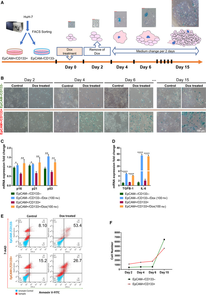
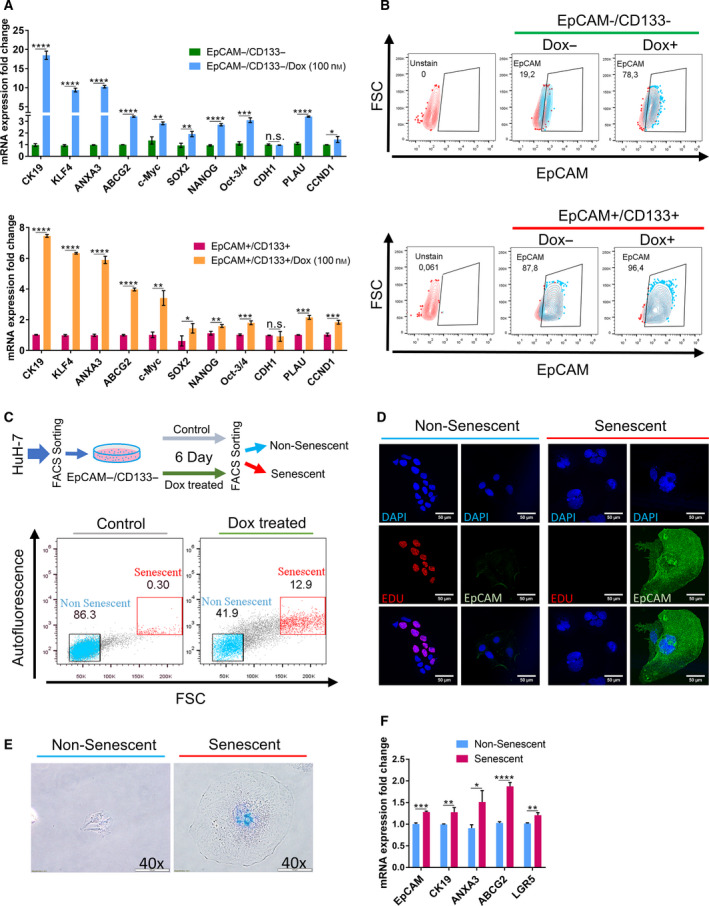
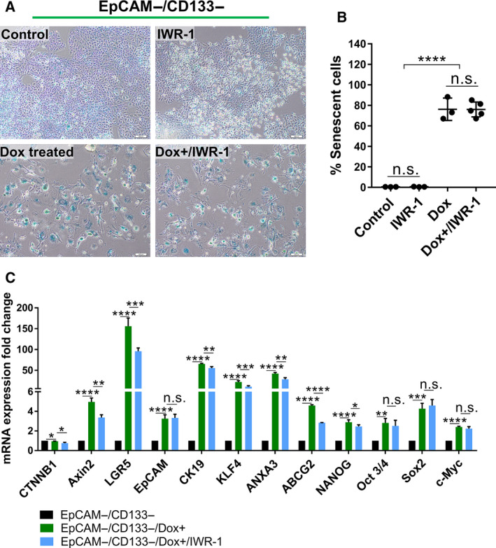
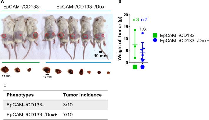
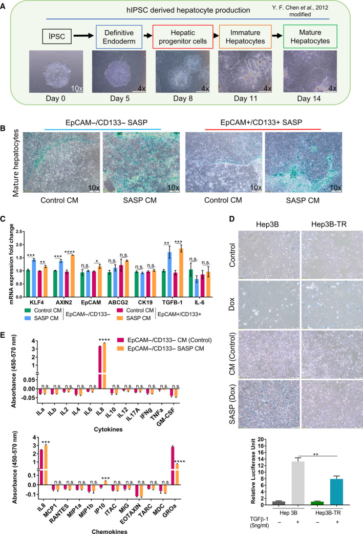
The therapeutic induction of senescence is a potential means to treat cancer, primarily acting through the induction of a persistent growth-arrested state in tumors. However, recent studies have indicated that therapy-induced senescence (TIS) in tumor cells allows for the prolonged survival of a subgroup of cells in a dormant state, with the potential to re-enter the cell cycle along with an increased stemness gene expression. Residual cells after TIS with increased cancer stem cell phenotype may have profound implications for tumor aggressiveness and disease recurrence. Herein, we investigated senescence-associated stemness in EpCAM+/CD133+ liver cancer stem cell and EpCAM-/CD133- nonstem cell populations in HuH7 cell line. We demonstrated that treatment with doxorubicin induces senescence in both cell populations, accompanied by a significant increase in the expression of reprogramming genes SOX2, KLF4, and c-MYC as well as liver stemness-related genes EpCAM, CK19, and ANXA3 and the multidrug resistance-related gene ABCG2. Moreover, doxorubicin treatment significantly increased EpCAM + population in nonstem cells indicating senescence-associated reprogramming of nonstem cell population. Also, Wnt/β-catenin target genes were increased in these cells, while inhibition of this signaling pathway decreased stem cell gene expression. Importantly, Dox-treated EpCAM-/CD133- nonstem cells had increased in vivo tumor-forming ability. In addition, when SASP-CM from Dox-treated cells were applied onto hİPSC-derived hepatocytes, senescence was induced in hepatocytes along with an increased expression of TGF-β, KLF4, and AXIN2. Importantly, SASP-CM was not able to induce senescence in Hep3B-TR cells, a derivative line rendered resistant to TGF-β signaling. Furthermore, ELISA experiments revealed that the SASP-CM of Dox-treated cells contain inflammatory cytokines IL8 and IP10. In summary, our findings further emphasize the importance of carefully dissecting the beneficial and detrimental aspects of prosenescence therapy in HCC and support the potential use of senolytic drugs in HCC treatment in order to eliminate adverse effects of TIS.

Keywords: EpCAM; WNT; cancer stem cell; hepatocellular carcinoma; therapy-induced senescence.


Conflict of interest statement

The authors declare no conflict of interest.

Figures

Fig. 1
Fig. 1
Analysis of induction of senescence and apoptosis in EpCAM+/CD133+ LCSCs and EpCAM‐/CD133‐ nonstem cells after Dox treatment. (A) The experimental workflow of therapy‐induced senescence model. (Scale bars: 50 μm) (B) Light microscope images of Dox‐treated cells after SA‐β‐gal staining on different days. (Scale bars: 100 μm) Expressional analysis of (C) senescence‐associated genes p16, p21, p53, and (D) SASP factors TGF‐β and IL‐6 by qPCR. (E) Flow cytometry analysis of Dox‐induced apoptosis using 7‐AAD/Annexin V staining. (F) Total cell number was counted and graphed for EpCAM+/CD133+ LCSCs and EpCAM−/CD133− nonstem cells on different days after Dox treatment. Two‐tailed unpaired Student’s t‐test was used to determine statistical significance. Data represent the average of at least three (n:5) independent experiments. P > 0.05 (n.s.), P ≤ 0.05 (*), P ≤ 0.01(**), P ≤ 0.001(***), P ≤ 0.0001(****). Error bars indicate standard deviation (SD).
Fig. 2
Fig. 2
Senescence‐associated stemness was analyzed in EpCAM+/CD133+ LCSCs and EpCAM−/CD133− nonstem cells. (A) Stemness‐related gene expression was analyzed by qPCR in EpCAM+/CD133+ LCSC and EpCAM−/CD133− nonstem cell subpopulations after Dox treatment at day 6. (B) Effect of Dox treatment on EpCAM + cell population was analyzed by flow cytometry using EpCAM‐FITC antibody. (C) Separation of nonsenescent and senescent cells from Dox‐treated EpCAM−/CD133− nonstem cells using size and autofluorescence differences via FACS. Enlarged cells with increased autofluorescence were isolated as senescent cells. (D) Immunofluorescence staining of isolated cells. The isolated senescent cells were negative for EdU staining while these senescent cells had high EpCAM staining. The nonsenescent cells were stained positively for EdU, however, did not show EpCAM expression. (Scale bars: 50 μm) (E) The validation of senescent and nonsenescent cells via SA‐βGal staining. (Scale bars: 100 μm) (F) qPCR analysis showed that senescent cells have increased expression of stemness‐related genes. Two‐tailed unpaired Student’s t‐test was used to determine statistical significance. Data represent the average of at least three (n:3) independent experiments. P > 0.05(n.s.), P ≤ 0.05 (*), P ≤ 0.01(**), P ≤ 0.001(***), P ≤ 0.0001(****). Error bars indicate standard deviation (SD).
Fig. 3
Fig. 3
The targeting of canonical Wnt/β‐catenin pathway reduces the expression of stemness‐related genes in EpCAM−/CD133− nonstem cells. (A) SA‐β‐gal staining of untreated and Dox‐treated EpCAM−/CD133− nonstem cells with and without IWR‐1 treatment. (Scale bars: 100 μm) (B) The number of senescent cells was graphed as percentage of the total cell number in all experimental groups. (C) The change in the expression of stemness‐related genes was analyzed by qPCR in all experimental groups. Two‐tailed unpaired Student’s t‐test was used to determine statistical significance. Data represent the average of at least three (n:3) independent experiments. P > 0.05 (n.s.), P ≤ 0.05 (*), P ≤ 0.01 (**), P ≤ 0.001 (***), P ≤ 0.0001 (****). Error bars indicate standard deviation (SD).
Fig. 4
Fig. 4
Dox‐treated EpCAM−/CD133− non‐LCSC cells have increased tumorigenicity. (A) NSG mice were subcutaneously injected with 10 000 untreated EpCAM−/CD133− cells or Dox‐treated EpCAM−/CD133− cells and then sacrificed after 41 days. (Scale bars: 10 mm). (B) Tumors were removed from the animals and weighed. (C) Dox‐treated EpCAM−/CD133− nonstem cells had significantly higher tumor incidence than control non‐LCSCs.
Fig. 5
Fig. 5
SASP‐CM of Dox‐treated EpCAM+/CD133+ LCSCs and EpCAM−/CD133− nonstem cells induce senescence in hepatocytes. (A) Production of hepatocytes from hiPSCs using our hepatic differentiation protocol. (Scale bars: 200 μm (Day 0) and 1 mm (Days 5–14) (B) The effect of SASP‐CM on hiPSCs derived hepatocytes. Cells were treated with SASP‐CM for 5 days with fresh SASP‐CM added every two days. (Scale bars: 200 μm) (C) The expression of stemness and inflammation‐related genes was analyzed by qPCR. (D) SA‐β‐gal staining of Hep3B and Hep3B‐TR cells incubated with SASP‐CM (top). Luciferase reporter assay using pSBE4‐Luc demonstrated reduced TGF‐β signaling in Hep3B‐TR cells in response to TGF‐β1 treatment (bottom). (Scale bars: 200 μm) (E) SASP‐CM from EpCAM−/CD133− control and 100 nm Dox‐treated EpCAM−/CD133− cells were analyzed using Human Common Chemokines Multi‐Analyte ELISArray Kit (Qiagen MEH‐009A) and Human Inflammatory Cytokines Multi‐Analyte ELISArray Kit (Qiagen MEH‐004A), respectively. Two‐tailed unpaired Student’s t‐test was used to determine statistical significance. Data represent the average of at least three (n:3) independent experiments. P > 0.05(n.s.), P ≤ 0.05 (*), P ≤ 0.01(**), P ≤ 0.001(***), P ≤ 0.0001(****). Error bars indicate standard deviation (SD).

References

    1. Losic B, Craig AJ, Villacorta‐Martin C, Martins‐Filho SN, Akers N, Chen X, Ahsen ME, von Felden J, Labgaa I, DʹAvola D et al. (2020) Intratumoral heterogeneity and clonal evolution in liver cancer. Nature Communications 11. 10.1038/s41467-019-14050-z - DOI - PMC - PubMed
    1. Friemel J, Rechsteiner M, Frick L, Böhm F, Struckmann K, Egger M, Moch H, Heikenwalder M & Weber A (2015) Intratumor heterogeneity in hepatocellular carcinoma. Clin Cancer Res 21, 1951–1961. - PubMed
    1. Dörr JR, Yu Y, Milanovic M, Beuster G, Zasada C, Däbritz JHM, Lisec J, Lenze D, Gerhardt A, Schleicher K et al. (2013) Synthetic lethal metabolic targeting of cellular senescence in cancer therapy. Nature 501, 421–425. 10.1038/nature12437 - DOI - PubMed
    1. Vilgelm AE, Johnson CA, Prasad N, Yang J, Chen SC, Ayers GD, Pawlikowski JS, Raman D, Sosman JA, Kelley M et al. (2016) Connecting the dots: therapy‐induced senescence and a tumor‐suppressive immune microenvironment. J Natl Cancer Inst 108. - PMC - PubMed
    1. Perez‐Amill L, Marzal B, Urbano‐Ispizua A, Juan M & Martín‐Antonio B (2018) CAR‐T cell therapy: a door is open to find innumerable possibilities of treatments for cancer patients. Turkish J Hematol. - PMC - PubMed

Publication types

MeSH terms