. 2021 Feb 19;16(2):389-396.
doi: 10.1021/acschembio.0c00937. Epub 2021 Feb 1.

A Bifunctional NAD+ for Profiling Poly-ADP-Ribosylation-Dependent Interacting Proteins

Affiliations

A Bifunctional NAD+ for Profiling Poly-ADP-Ribosylation-Dependent Interacting Proteins

Albert T Lam et al. ACS Chem Biol. .

Abstract

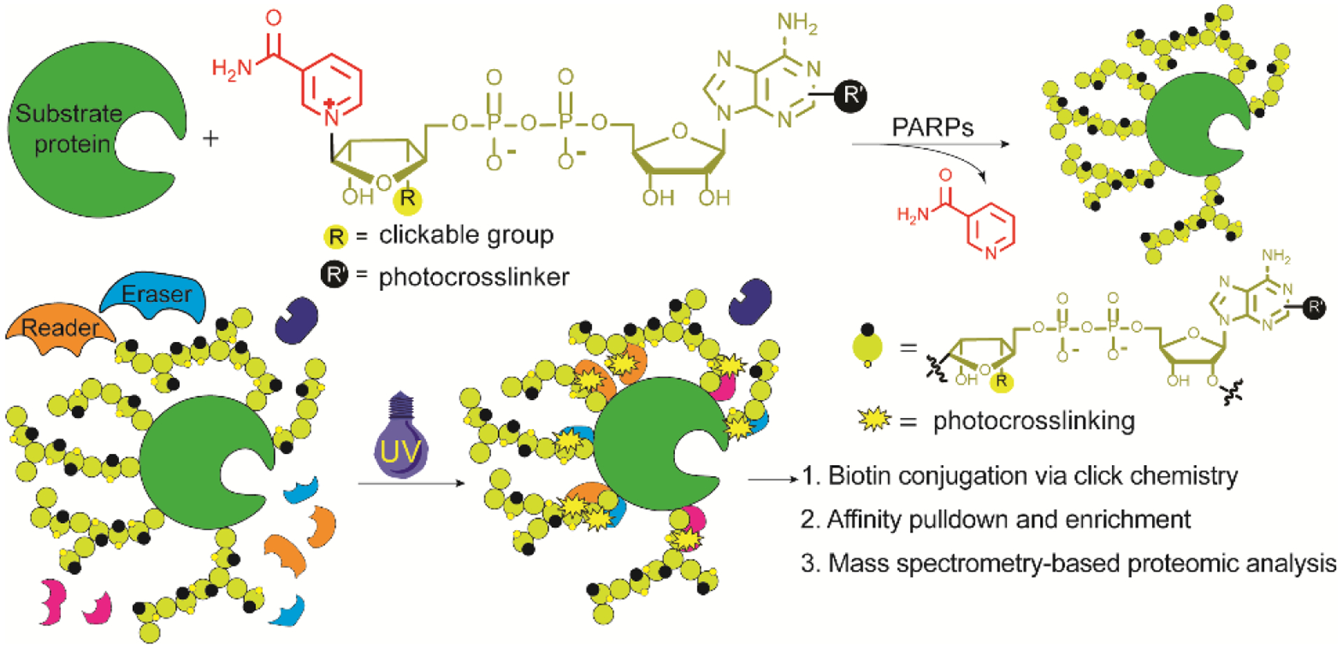
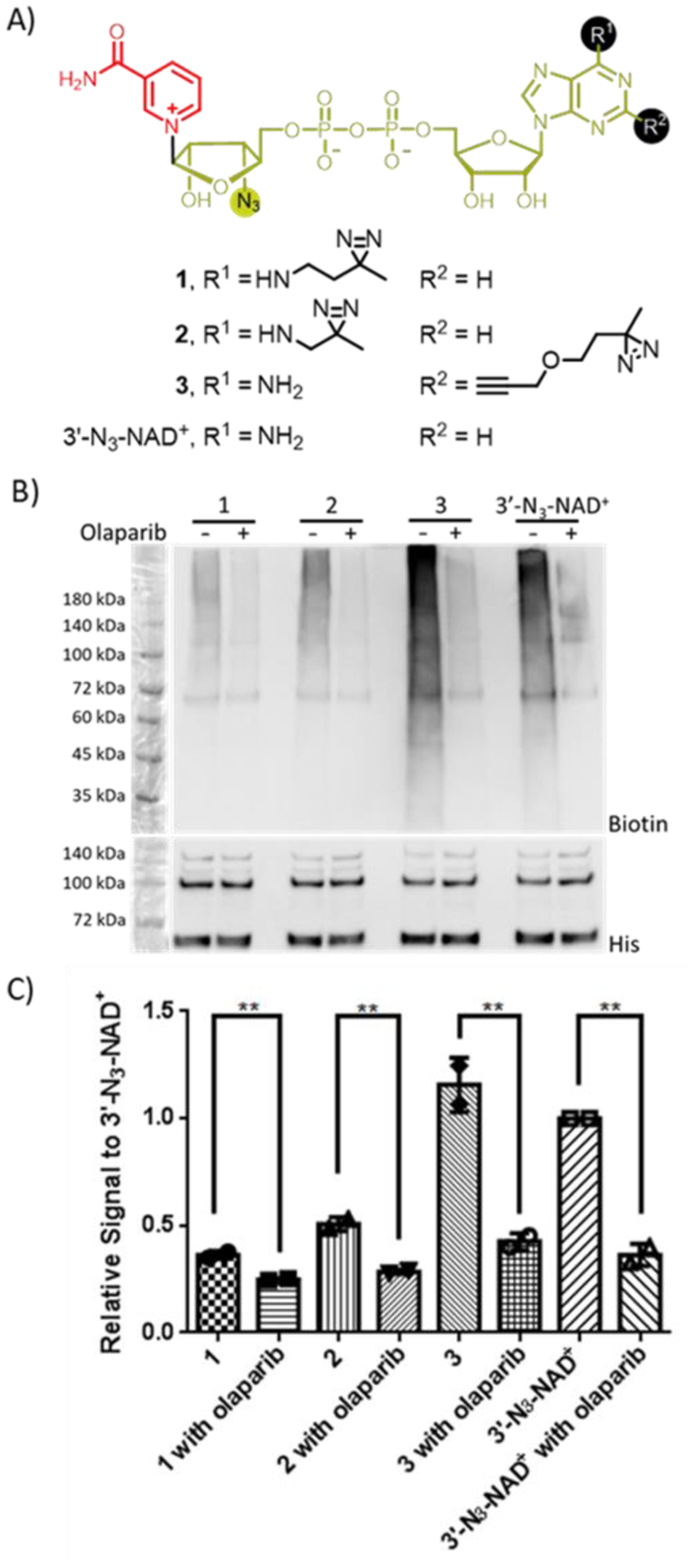
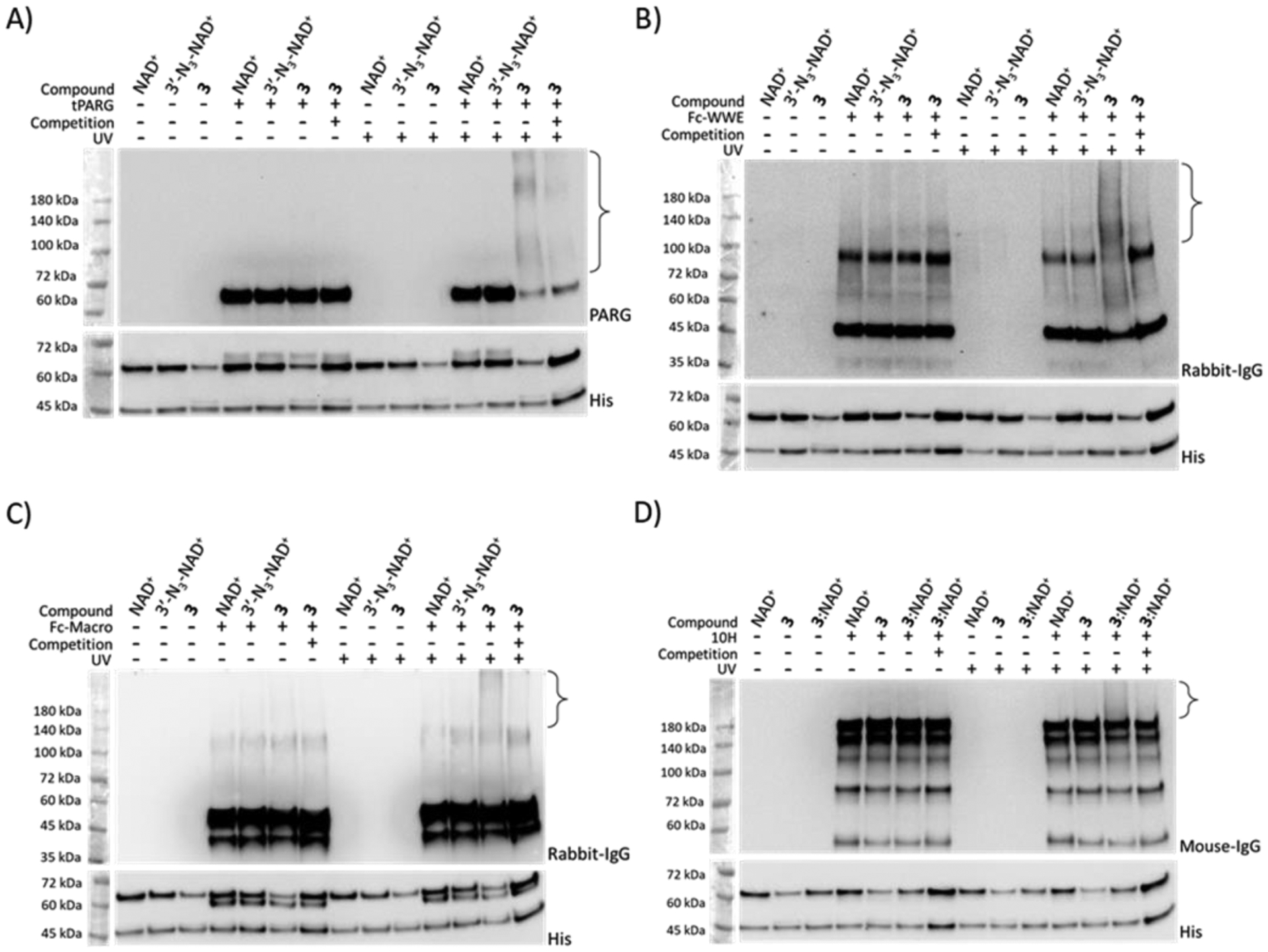
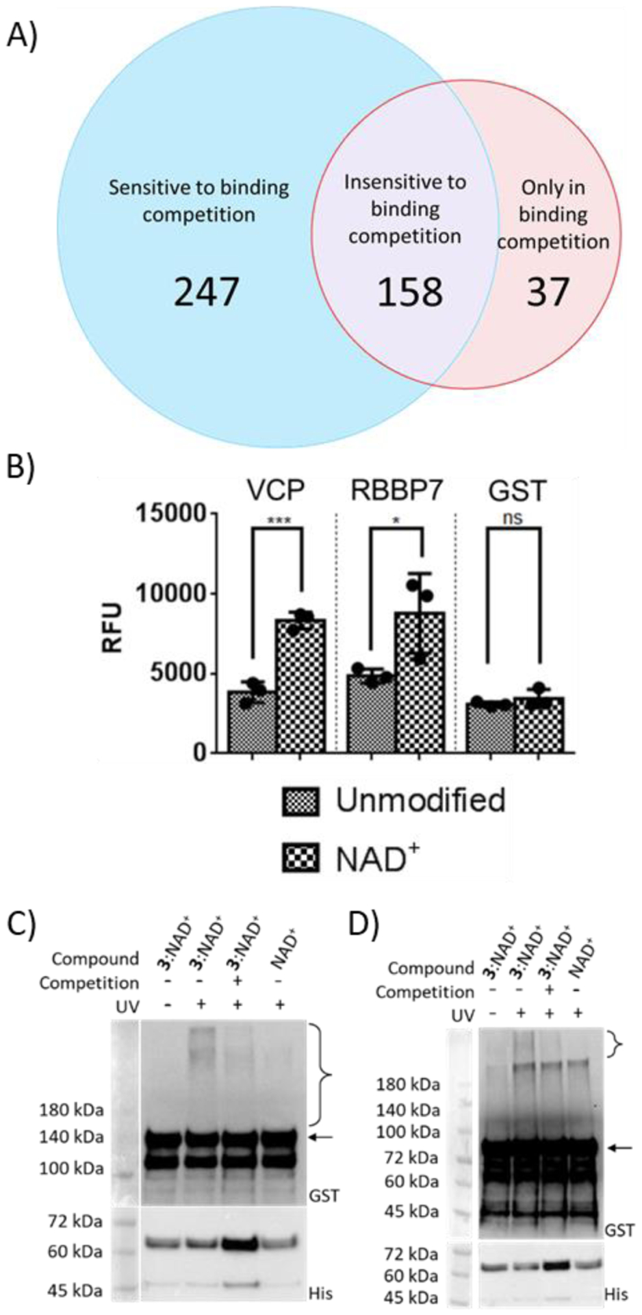
Protein poly-ADP-ribosylation (PARylation) is a heterogeneous and dynamic post-translational modification regulated by various writers, readers, and erasers. It participates in a variety of biological events and is involved in many human diseases. Currently, tools and technologies have yet to be developed for unambiguously defining readers and erasers of individual PARylated proteins or cognate PARylated proteins for known readers and erasers. Here, we report the generation of a bifunctional nicotinamide adenine dinucleotide (NAD+) characterized by diazirine-modified adenine and clickable ribose. By serving as an excellent substrate for poly-ADP-ribose polymerase 1 (PARP1)-catalyzed PARylation, the generated bifunctional NAD+ enables photo-cross-linking and enrichment of PARylation-dependent interacting proteins for proteomic identification. This bifunctional NAD+ provides an important tool for mapping cellular interaction networks centered on protein PARylation, which are essential for elucidating the roles of PARylation-based signals or activities in physiological and pathophysiological processes.


Conflict of interest statement

Conflict of Interest Disclosure

The authors declare no competing financial interests.

Figures

Figure 1.
Figure 1.
Profiling PARylation-dependent interacting proteins by a bifunctional NAD+.
Figure 2.
Figure 2.
Substrate activities of NAD+ analogues for human PARP1. (A) Chemical structures of NAD+ analogues. (B) Immunoblot analysis of auto-PARylation of PARP1 with NAD+ analogues using a streptavidin-HRP conjugate following biotin conjugation via click chemistry. Auto-PARylation reactions were performed in the absence or presence of the PARP inhibitor olaparib. Lower panel: PARP1 loading controls detected using an anti-His6 antibody. The observed bands between 60 and 72 kDa were proteolyzed PARP1 formed during auto-modification reactions. (C) Relative densitometric analysis of PARylation levels. Error bars represent standard deviation of two replicates.
Figure 3.
Figure 3.
Photocrosslinking of automodified PARP1 with model interacting proteins. PARP1 PARylated by NAD+, 3′-N3-NAD+, 3, or a mixture of 3:NAD+ at a 1:1 molar ratio was incubated with (A) tPARG, (B) Fc-WWE, (C) Fc-Macro, and (D) antibody 10H in the absence or presence of 365-nm UV irradiation and automodified PARP1 by NAD+ for binding competition, followed by immunoblot analysis using an anti-PARG antibody for (A) tPARG, an anti-rabbit IgG antibody for (B) Fc-WWE and (C) Fc-Macro, and an anti-mouse IgG antibody for (D) antibody 10H. Lower panels: PARP1 loading controls detected using an anti-His6 antibody. The observed bands between 45 and 72 kDa were proteolyzed PARP1 formed during auto-modification reactions. Brackets indicate regions of crosslinked protein complexes. Full-sized anti-His6 immunoblots are shown in Figure S5.
Figure 4.
Figure 4.
Verification of novel PARylation-dependent interacting proteins. (A) Venn diagram of protein hits identified from photocrosslinking in cell lysates. (B) Binding of GST-VCP and GST-RBBP7 for unmodified PARP1 and automodified PARP1 by NAD+. Unmodified PARP1 and NAD+-modified PARP1 were coated on ELISA plates for binding analysis with two identified protein hits. ns: not significant, *P < 0.05, *** P < 0.001 by one-tailed unpaired t-test. Error bars represent standard deviation of three replicates. RFU: relative fluorescence unit. (C) and (D) Photocrosslinking of automodified PARP1 with (C) VCP and (D) RBBP7. PARP1 PARylated by NAD+ or a mixture of 3:NAD+ (molar ratio 1:1) was incubated with GST-VCP or GST-RBBP7 without and with 365-nm UV and NAD+-modified PARP1 for binding competition, followed by immunoblot analysis using an anti-GST antibody. Expected sizes for VCP and RBBP7 are indicated by the arrow for (C) and (D) respectively. Lower panels: PARP1 loading controls detected using an anti-His6 antibody. The observed bands between 45 and 72 kDa were proteolyzed PARP1 formed during auto-modification reactions. Brackets indicate regions of crosslinked protein complexes.

References

    1. Andrabi SA, Kang HC, Haince JF, Lee YI, Zhang J, Chi Z, West AB, Koehler RC, Poirier GG, Dawson TM, and Dawson VL (2011) Iduna protects the brain from glutamate excitotoxicity and stroke by interfering with poly(ADP-ribose) polymer-induced cell death, Nat. Med 17, 692–699. - PMC - PubMed
    1. Han W, Li X, and Fu X (2011) The macro domain protein family: structure, functions, and their potential therapeutic implications, Mutat. Res 727, 86–103. - PMC - PubMed
    1. Kalisch T, Ame JC, Dantzer F, and Schreiber V (2012) New readers and interpretations of poly(ADP-ribosyl)ation, Trends Biochem. Sci 37, 381–390. - PMC - PubMed
    1. Pleschke JM, Kleczkowska HE, Strohm M, and Althaus FR (2000) Poly(ADP-ribose) binds to specific domains in DNA damage checkpoint proteins, J. Biol. Chem 275, 40974–40980. - PubMed
    1. Rack JG, Perina D, and Ahel I (2016) Macrodomains: Structure, Function, Evolution, and Catalytic Activities, Annu. Rev. Biochem 85, 431–454. - PubMed

Publication types

MeSH terms

LinkOut - more resources