. 2021 Apr 2;433(7):166833.
doi: 10.1016/j.jmb.2021.166833. Epub 2021 Jan 30.

Clusters of a Few Bound Cofilins Sever Actin Filaments

Affiliations

Clusters of a Few Bound Cofilins Sever Actin Filaments

Jeffrey P Bibeau et al. J Mol Biol. .

Abstract

Cofilin is an essential actin filament severing protein that accelerates the assembly dynamics and turnover of actin networks by increasing the number of filament ends where subunits add and dissociate. It binds filament subunits stoichiometrically and cooperatively, forming clusters of contiguously-bound cofilin at sub-saturating occupancies. Filaments partially occupied with cofilin sever at boundaries between bare and cofilin-decorated segments. Imaging studies concluded that bound clusters must reach a critical size (Cc) of 13-100 cofilins to sever filaments. In contrast, structural and modeling studies suggest that a few or even a single cofilin can sever filaments, possibly with different severing rate constants. How clusters grow through the cooperative incorporation of additional cofilin molecules, specifically if they elongate asymmetrically or uniformly from both ends and if they are modulated by filament shape and external force, also lacks consensus. Here, using hydrodynamic flow to visualize individual actin filaments with TIRF microscopy, we found that neither flow-induced filament bending, tension, nor surface attachment conditions substantially affected the kinetics of cofilin binding to actin filaments. Clusters of bound cofilin preferentially extended toward filament pointed ends and displayed severing competency at small sizes (Cc < 3), with no detectable severing dependence on cluster size. These data support models in which small clusters of cofilin introduce local, but asymmetric, structural changes in actin filaments that promote filament severing with a rate constant that depends weakly on the size of the cluster.

Keywords: curvature; fluorescence; kinetics; microfluidics; tension.


Conflict of interest statement

Declaration of Competing Interest The authors declare no competing interest.

Figures

Figure 1.
Figure 1.
Cofilin association, but not dissociation, is independent of applied flow force and filament bending. (A) Example association experiments where 200 nM cofilin is flown over pointed end tethered actin filaments at 500 (bottom) and 5 (top) μL min−1 flow. Alexa 647-actin filaments are colored magenta and Alexa 488-cofilin is colored cyan. Scale bar is 2 μm. (B) Time courses of normalized actin filament-bound cofilin fluorescence intensity during association in (A) (see Methods). n = 10 and 14 for the 500 (black) and 5 (blue) μL min-1 flow rates, respectively. Only filaments without severing events were analyzed. Uncertainty bars represent the standard error of the mean intensity. Average filament lengths are 9.7 and 7.7 μm for high and low flow, respectively. (C) Cumulative distribution function of the observable cluster formation positions from filament free ends. Positions were determined 15 s after addition of 1 μM cofilin. A total of 19 filaments with 230 clusters were used. Traces correspond to filaments with comparable lengths (blue = 8–11 mm, black = 11–14 mm, gray = 14–17 μm, and green = 17–20 μm). (D) Time courses of competitive cofilin dissociation via stopped flow and fluorescence microscopy. Dissociation of Alexa-488 cofilin was measured under 500 μL min−1 flow (black circles) and 5 μL min−1 flow (blue circles) with 1 μM unlabeled cofilin in the flow buffer. n = 19 and 18 filaments, respectively. Uncertainty bars represent the standard error of the mean. Dissociation of Oregon green-labeled cofilin was measured via fluorescence change with stopped flow (continuous blue line) in solution by competition with unlabeled cofilin (see Methods). All time courses were fit to single exponentials (red lines) yielding cooperative dissociation rate constants of 2.3 × 10⁻⁴ s−1 (95% CI 2–2.5 × 10⁻⁴) under 500 μL min−1 flow, 7.1 × 10⁻⁴ s−1 (95% CI 5.8–8.4 × 10⁻⁴) under 5 μL min−1 flow, and 1.6 × 10⁻³ s−1 (95% CI 1.5–1.6 × 10⁻³) under stopped flow.
Figure 2.
Figure 2.
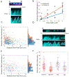
Cofilin forms clusters that preferentially elongate toward the filament pointed end. (A) Cofilin association on 15% Alexa-647-labeled filaments (top). Kymographs of association (middle) and manual tracing of cofilin extension (bottom). (B) Cofilin cluster extension rates towards the barbed (blue) and pointed ends (orange) as measured during association with kymographs. n > 20 clusters for each condition. Slopes of best fit lines yield cluster elongation rates of 3.3 (95% CI 1.8–4.9) and 6.3 (95% CI 4.8–7.8) μM−1 s−1 towards the barded and pointed ends, respectively. Uncertainty bars represent the standard error of the mean for each point. Significance was determined with Dunn’s multiple comparison test. The * indicates p < 0.05. C) Cofilin cluster extension rates towards the barbed (blue) and pointed ends (orange) of Alexa-647-labeled filaments as a function of the distance from the center of the cluster from the barbed end. The scatter along the x-axis is due to the filament length distribution and not tension. D) Cofilin association on unlabeled filaments (top). Kymographs of association (middle) and manual tracking of cofilin extension (bottom). E) Cofilin cluster extension rates towards the barbed (blue) and pointed ends (orange) on unlabeled filaments as a function of the distance of the center of the cluster from the barbed end. F) Box plot of cofilin extension rates during 200 nM association on unlabeled (black points) and Alexa-647 (red points) labeled filaments. The red line represents the median, the blue box indicates the 25th and 75th percentiles, whiskers indicate the data spread excluding outliers, and plus signs indicate outliers. Points were classified as outliers if they were greater than q3 + w × (q3 − q1) or less than q1 − w × (q3 − q1). Here q1, is the first quartile, q2 is the second quartile, and w is the maximum whisker which extends to the points closest to ±2.7σ.
Figure 3.
Figure 3.
A few cofilins can sever a filament. (A) Photobleaching of a cofilin cluster (black circles) and the predicted bleach steps (red line) (see Methods). (B) Histogram of model fit bleach steps for all the clusters measured during rapid photobleaching on filaments completely adhered to the surface with biotin. The red line represents the best fit of the data to a mixture of Gaussians. The means for the two prominent peaks are 70 and 160. The inset depicts the ensemble bleaching rate for all the clusters. The red line represents the best fit of the data to a single exponential yielding a rate constant of 6.4 × 10⁻³ frame s−1 which is a combination of bleaching and dissociation. (C) Filament severing during 50 (left) and 200 (right) nM cofilin association. Filaments were attached to the surface near their pointed ends. The middle image is an intensity rescaling of the leftmost image to better visualize the clusters. (D) Size of bound cofilin clusters (measured via fluorescence) prior to observed filament severing events. During 50 nM cofilin severing experiments, filaments were biotinylated throughout their length or tethered at their pointed end. During 200 nM cofilin severing experiments, filaments were only tethered at their pointed ends.
Figure 4.
Figure 4.
Cofilin boundary severing is not strongly dependent on cluster size: (A and B) The lifetime of unsevered boundaries at the pointed end (A) and barbed end (B) of cofilin clusters with 50 nM (black) and 200 nM (blue) free cofilin in solution. 95% confidence bounds are indicated by shaded regions. Kaplan Meir corrections were applied to account for boundaries that were not severed or lost.

Similar articles

Cited by

References

    1. De La Cruz EM, (2009). How cofilin severs an actin filament. Biophys. Rev, 1, 51–59. - PMC - PubMed
    1. Kanellos G, Frame MC, (2016). Cellular functions of the ADF/cofilin family at a glance. J. Cell Sci, 129, 3211–3218. - PubMed
    1. McGough A, Pope B, Chiu W, Weeds A, (1997). Cofilin changes the twist of F-actin: implications for actin filament dynamics and cellular function. J. Cell Biol, 138, 771–781. - PMC - PubMed
    1. De La Cruz EM, (2005). Cofilin binding to muscle and non-muscle actin filaments: isoform-dependent cooperative interactions. J. Mol. Biol, 346, 557–564. - PubMed
    1. McCullough BR, Grintsevich EE, Chen CK, Kang HR, Hutchison AL, Henn A, et al., (2011). Cofilin-linked changes in actin filament flexibility promote severing. Biophys. J, 101, 151–159. - PMC - PubMed

Publication types

MeSH terms

LinkOut - more resources