

mAb Das-1 identifies pancreatic ductal adenocarcinoma and high-grade pancreatic intraepithelial neoplasia with high accuracy
- PMID: 33524436
- PMCID: PMC8817765
- DOI: 10.1016/j.humpath.2021.01.003
mAb Das-1 identifies pancreatic ductal adenocarcinoma and high-grade pancreatic intraepithelial neoplasia with high accuracy
Abstract
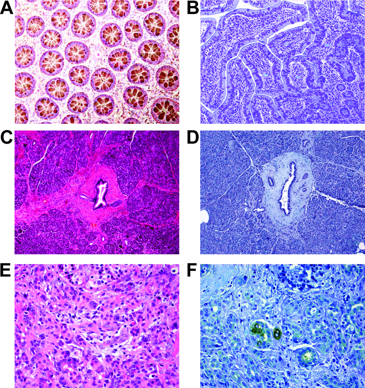
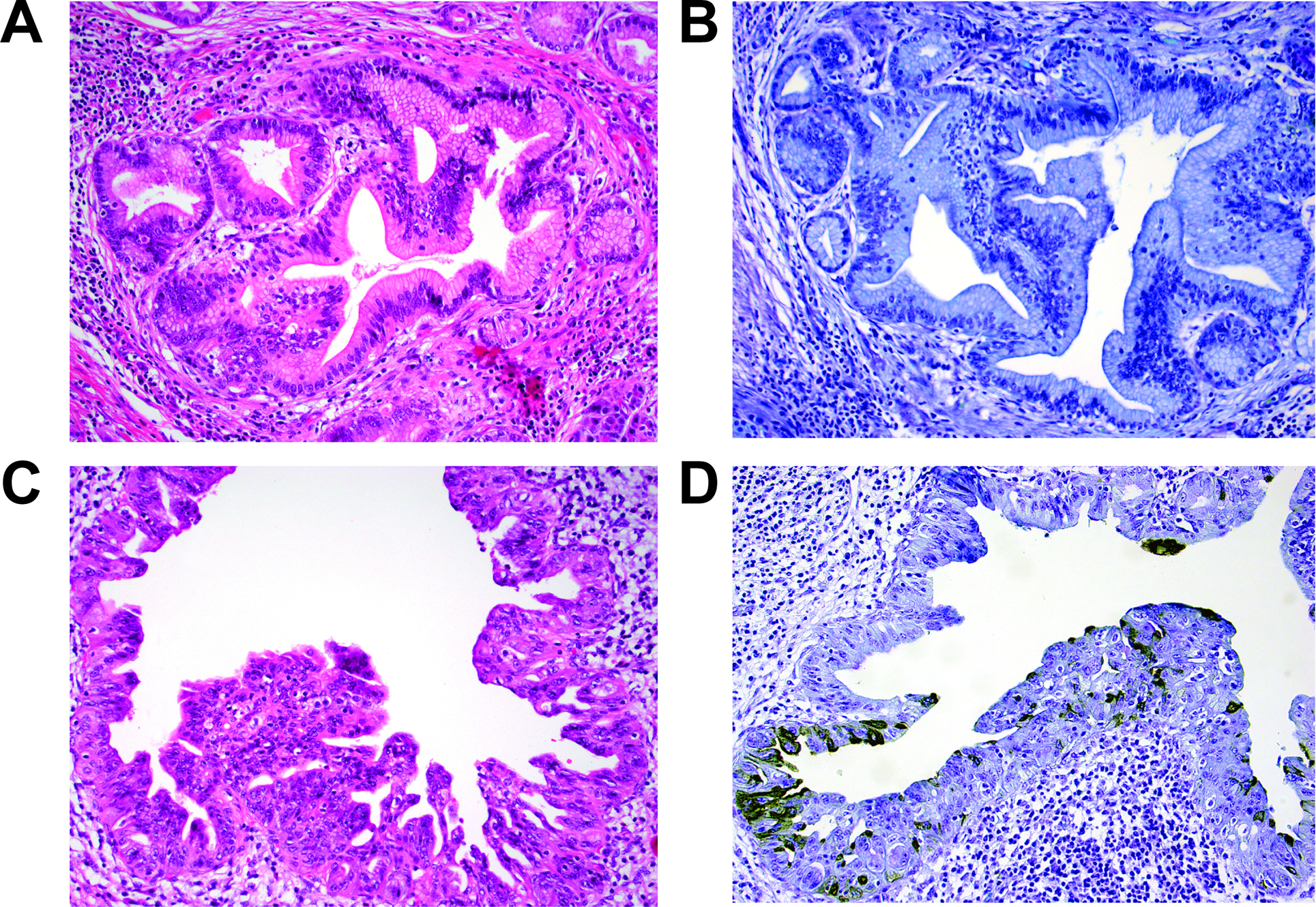
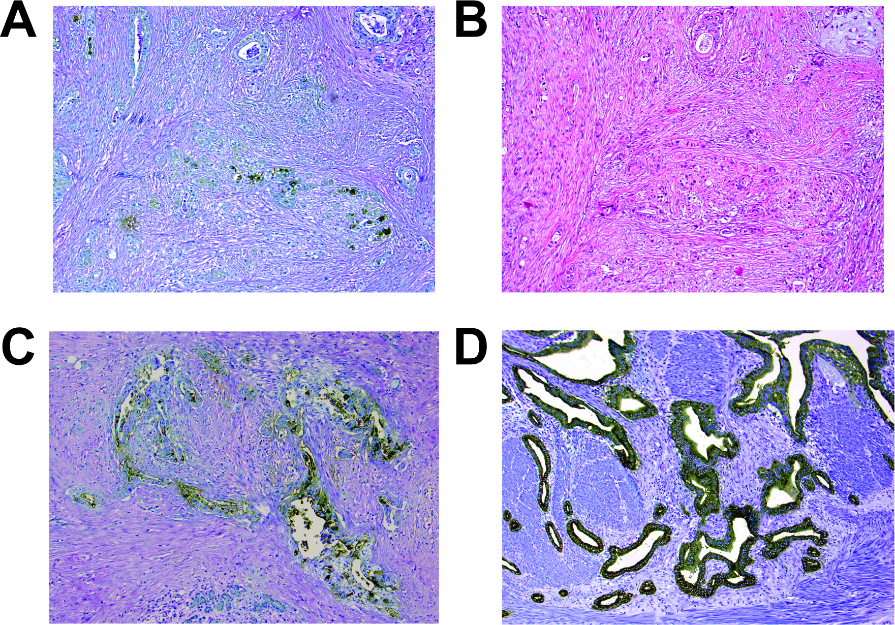
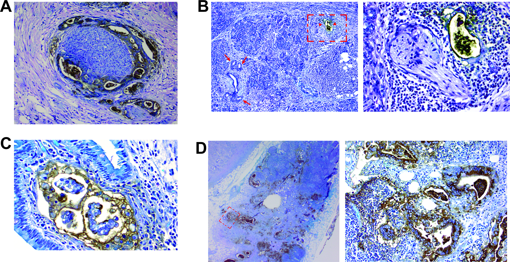
Pancreatic intraepithelial neoplasia (PanIN) is a microscopic precursor lesion to pancreatic ductal adenocarcinoma (PDAC); however, there are few biomarkers that segregate high-grade PanIN/PDAC from low-grade PanIN lesions. mAb Das-1 is a monoclonal antibody against a colonic epithelial antigen that is reactive to premalignant conditions of the upper gastrointestinal tract including Barrett's esophagus, incomplete-type gastric intestinal metaplasia, and intraductal papillary mucinous neoplasm of the pancreas at high risk of malignancy. We sought to examine a role for Das-1 expression in differentiating high-grade PanIN/PDAC from low-grade PanIN lesions. We examined surgical specimens from 86 patients and 2 autopsied pancreata (74 with and 14 without PDAC) with 107 distinct PanIN lesions, 74 PDAC cases, and 32 associated lymph node metastases, with internal controls of normal pancreatic ducts observed in 56 cases. All of the normal pancreatic duct controls (0/56) and low-grade PanIN (0/95) lesions were nonreactive to Das-1. Das-1 expression among high-grade PanIN (7/12, 58%), PDAC (55/74, 74%), and lymph node metastasis (21/32, 66%) cases was significantly higher (p < 0.0001). Clinicopathologically, Das-1 reactivity was significantly correlated with nodal metastasis (p = 0.021). Overall, the sensitivity, specificity, and accuracy of Das-1 in segregating high-grade PanIN/PDAC from low-grade PanIN lesions and normal ducts were 72%, 100%, and 90%, respectively. Thus, mAb Das-1 reacts with high specificity with high-grade PanIN and PDAC and may help in preoperative diagnosis and/or clinical risk stratification.
Keywords: Biomarker; CEP; Pancreatic adenocarcinoma; Pancreatic intraepithelial neoplasia (PanIN); mAb Das-1.
Copyright © 2021 Elsevier Inc. All rights reserved.
Conflict of interest statement
DECLARATION OF INTEREST
A patent for the use of mAb Das-1 in the detection of precancerous lesions of the esophagus has been awarded (KMD) and a patent for its possible use in the detection of pancreatic cancer is currently pending (KKD, MMK, KMD). These patents have not been licensed and these authors hold no commercial interests at this time.
Figures
References
-
- Rahib L, Smith BD, Aizenberg R, Rosenzweig AB, Fleshman JM, Matrisian LM. Projecting cancer incidence and deaths to 2030: the unexpected burden of thyroid, liver, and pancreas cancers in the United States. Cancer research 2014; 74, 2913–2921. - PubMed
-
- Siegel R, Ward E, Brawley O, Jemal A. Cancer statistics, 2011. CA: A Cancer Journal for Clinicians 2011; 61, 212–236. - PubMed
-
- Hruban RH, Goggins M, Parsons J, Kern SE. Progression model for pancreatic cancer. Clin Cancer Res 2000; 6, 2969–2972. - PubMed
-
- Hruban RH, Takaori K, Klimstra DS, Adsay NV, Albores-Saavedra J, Biankin AV, Biankin SA, Compton C, Fukushima N, Furukawa T, Goggins M, Kato Y, Kloppel G, Longnecker DS, Luttges J, Maitra A, Offerhaus GJ, Shimizu M, Yonezawa S. An illustrated consensus on the classification of pancreatic intraepithelial neoplasia and intraductal papillary mucinous neoplasms. Am J Surg Pathol 2004; 28, 977–987. - PubMed
-
- Basturk O, Hong SM, Wood LD, Adsay NV, Albores-Saavedra J, Biankin AV, Brosens LA, Fukushima N, Goggins M, Hruban RH, Kato Y, Klimstra DS, Kloppel G, Krasinskas A, Longnecker DS, Matthaei H, Offerhaus GJ, Shimizu M, Takaori K, Terris B, Yachida S, Esposito I, Furukawa T, Baltimore Consensus M. A Revised Classification System and Recommendations From the Baltimore Consensus Meeting for Neoplastic Precursor Lesions in the Pancreas. The American journal of surgical pathology 2015; 39, 1730–1741. - PMC - PubMed
Publication types
MeSH terms
Substances
Grants and funding
LinkOut - more resources
Full Text Sources
Other Literature Sources
Medical
